Isolated Double-Chambered Right Ventricle in a Young Adult
- Affiliations
-
- 1Department of Internal Medicine, Daegu Fatima Hospital, Daegu, Korea. tangul8845@hanmail.net
- KMID: 2297935
- DOI: http://doi.org/10.4070/kcj.2011.41.5.272
Abstract
- Double-chambered right ventricle (DCRV) is a rare congenital heart disorder in which the right ventricle is divided by an anomalous muscle bundle into a high pressure inlet portion and a low pressure outlet portion. We report a case of isolated DCRV without symptoms in adulthood, diagnosed through echocardiography, cardiac catheterization and cardiac magnetic resonance imaging.
Keyword
MeSH Terms
Figure
-
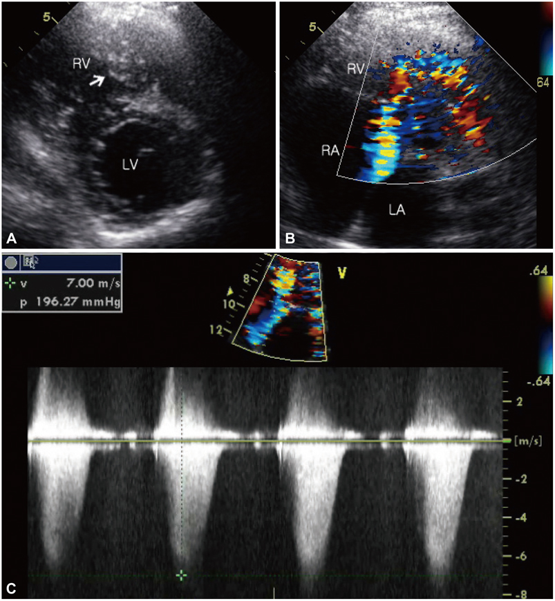
Fig. 1 Double-chambered right ventricle demonstrated by transthrorasic echocardiography. A: two-dimensional echocardiography showing a stenotic mid-right ventricle with an anomalous muscle bundle (arrow). B: turbulent Doppler color flow with mosaic pattern. C: continuous-wave Doppler showing tricuspid regurgitation between the right atrium and right ventricle and indicating the flow acceleration and pressure gradient. RV: right ventricle, LV: left ventricle, RA: right atrium.

Fig. 2 Transesophageal echocardiography showing anomalous muscular bundles (arrow) dividing the right ventricle into two parts. RV: right ventricle, LV: left ventricle, RA: right atrium, LA: right atrium.
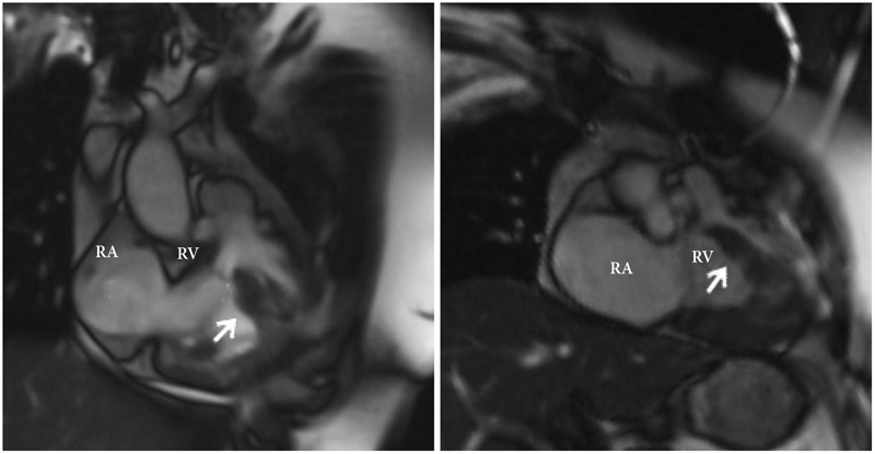
Fig. 3 Two-dimensional cine MRI showing anomalous muscle bundle (arrow). RV: right ventricle, RA: right atrium.
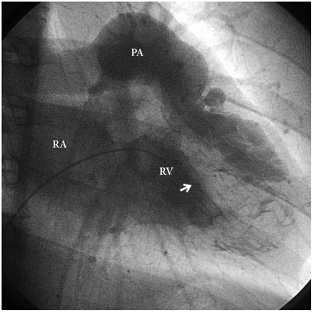
Fig. 4 Right ventriculogram demonstrating a severe hypertrophied muscle bundle (arrow). RV: right ventricle, RA: right atrium, PA: pulmonary artery.

Fig. 5 Cardiac catheterization showing systolic pressure gradient between RVIT and RVOT. RVIT: right ventricular inlet tract, RVOT: right ventricular outlet tract.
Reference
-
1. Cil E, Saraclar M, Özkutlu S, et al. Double-chambered right ventricle: experience with 52 cases. Int J Cardiol. 1995. 50:19–29.2. Kim CJ, Chai IH, Koh KK, et al. Double chambered right ventricle in adult and adolescence. Korean Circ J. 1990. 20:248–255.3. Nishimura RA, Miller FA Jr, Callahan MJ, Benassi RC, Seward JB, Tajik AJ. Doppler echocardiography: theory, instrumentation, technique, and application. Mayo Clin Proc. 1985. 60:321–343.4. Galiuto L, O'Leary PW, Seward JB. Double-chambered right ventricle: echocardiographic feature. J Am Soc Echocardiogr. 1996. 9:300–305.5. Choi YJ, Park SW. Characteristics of double-chambered right ventricle in adult patients. Korean J Intern Med. 2010. 25:147–153.6. Hoffman P, Wojcik AW, Rozanski J, et al. The role of echocardiography in diagnosing double chambered right ventricle in adult. Heart. 2004. 90:789–793.7. Galal O, Al-Halees Z, Solymar L, et al. Double-chambered right ventricle in 73 patients: spectrum of the disease and surgical results of transartial repair. Can J Cardiol. 2000. 16:167–174.8. Chang RY, Kou CH, Rim RS, Chou YS, Tsai CH. Transesophageal echocardiographic image of double-chambered right ventricle. J Am Soc Echocardiogr. 1996. 9:347–352.9. Sato Y, Matsumoto N, Matsuo S, et al. Double-chambered right ventricle: depiction at three-dimensional whole heart magnetic resonance imaging. Int J Cardiol. 2007. 119:e14–e16.10. McElhinney DB, Chatterjee KM, Reddy VM. Double-chambered right ventricle presenting in adulthood. Ann Thorac Surg. 2000. 70:124–127.11. Kim J, Park TI, An SG, et al. Electrical storm late after surgery for a double-chambered right ventricle, aortic regurgitation and a ventricular septal defect: a case of successful catheter ablation. Korean Circ J. 2008. 38:60–65.12. Hachiro Y, Takagi N, Koyanagi T, Morikawa M, Abe T. Repair of double-chambered right ventricle: surgical results and long-term follow-up. Ann Thorac Surg. 2001. 72:1520–1522.13. Yang SM, Chung WJ, Oh KJ, Kim MJ, Kim MK, Ahn TH. Two cases of double-chambered right ventricle without other congenital cardiac anomalies. J Korean Soc Echocardiogr. 2005. 13:37–41.14. Vieillard-Baron A. Assessment of right ventricular function. Curr Opin Crit Care. 2009. 15:254–260.
- Full Text Links
-
- Actions
-
Cited
- CITED
-
- Close
- Share
-
- Similar articles
-
- A Case of Double Chambered Right Ventricle with Congenital Right Ventricular True Diverticulum
- Two Cases of Double-Chambered Right Ventricle without Other Congenital Cardiac Anomalies
- Two Cases of Double-Chambered Right Ventricle by Abnomal Muscle Bundles
- Double Chambered Right Ventricle with Ventricular Septal Defect in Adults: Case Series and Review of the Literature
- Noonan Syndrome with Double-Chambered Right Ventricle and Atrial Septal Defect: 1 Case Report
