Lateralization of Hypoglycemic Encephalopathy: Evidence of a Mechanism of Selective Vulnerability
- Affiliations
-
- 1Department of Neurology, Kangwon National University College of Medicine, Chuncheon, Korea.
- 2Department of Internal Medicine, Kangwon National University College of Medicine, Chuncheon, Korea.
- 3Department of Radiology, Kangwon National University College of Medicine, Chuncheon, Korea. samskim@kangwon.ac.kr
- 4Medical Science and Engineering Research Center for Bioreaction to Reactive Oxygen Species, Kangwon National University College of Medicine, Chuncheon, Korea.
- KMID: 2287642
- DOI: http://doi.org/10.3988/jcn.2010.6.2.104
Abstract
- BACKGROUND
One of the characteristics of hypoglycemic encephalopathy (HE) is selective vulnerability of different brain regions.
CASE REPORT
We observed a patient with unilateral HE affecting the right internal capsule and the subcortical white matter. The patient had a preexisting stroke in the opposite hemisphere. The hemisphere that was affected by HE exhibited greater regional blood flow (single positron-emission tomography) and higher fractional anisotropy (diffusion-tensor imaging) than the unaffected hemisphere.
CONCLUSIONS
This case suggests that the degree of metabolism required to maintain the function of brain structures and neuronal integrity is an important factor determining the selective vulnerability in HE.
Keyword
Figure
-
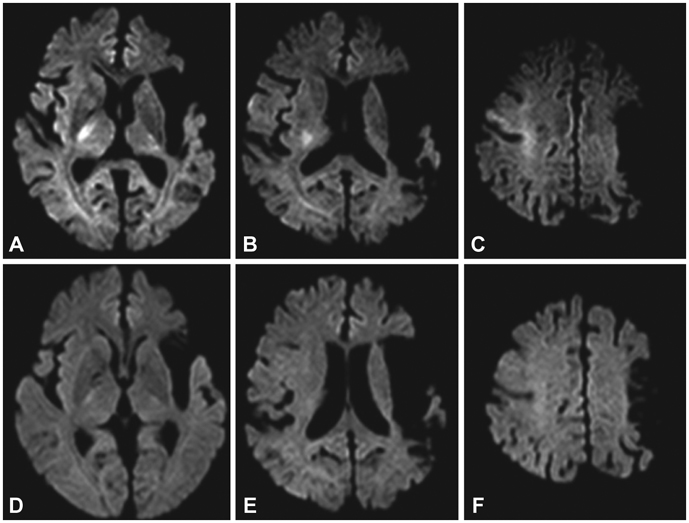
Fig. 1 The initial and follow-up diffusion-weighted images of the patient. The initial images (A-C) demonstrate lateralizing high-signal-intensity lesions in the right internal capsule, periventricular WM, and frontal cortex. On the follow-up images (D-F), the initial hyperintense lesions disappear. WM: white matter.
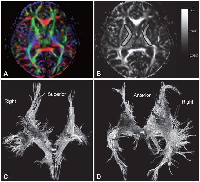
Fig. 2 The diffusion tensor images of the patient. A: Color-coded directional map of the FA demonstrates the relatively lower integrity in the left internal capsule. B: The ROIs positioned on the internal capsule that were used to calculate the FA values. C and D: Reconstructed three-dimensional tractography shows the relatively scanty descending fibers in the left hemisphere. The gray scale bar represents the FA value. FA: fractional anisotropy, ROIs: regions of interest.
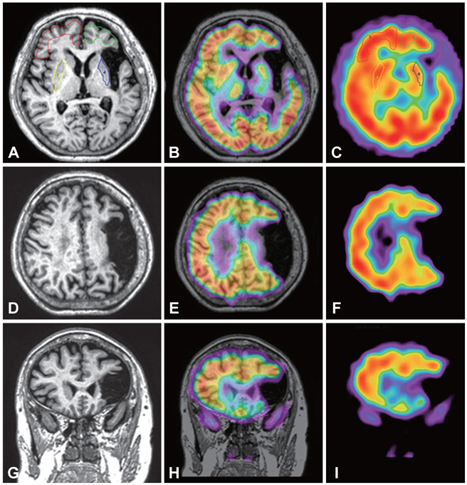
Fig. 3 T1 MR images (A, D and G), coregistered MRI to SPECT (B, E and H), and SPECT (C, F and I) demonstrate the relatively lower rCBF in the left hemisphere. The four ROIs (red, green, yellow, and blue lines in A and C) are positioned to calculate the asymmetric index. rCBF: regional cerebral blood flow, ROIs: regions of interest.
Reference
-
1. Lo L, Tan AC, Umapathi T, Lim CC. Diffusion-weighted MR imaging in early diagnosis and prognosis of hypoglycemia. AJNR Am J Neuroradiol. 2006. 27:1222–1224.2. Aoki T, Sato T, Hasegawa K, Ishizaki R, Saiki M. Reversible hyperintensity lesion on diffusion-weighted MRI in hypoglycemic coma. Neurology. 2004. 63:392–393.
Article3. Chan R, Erbay S, Oljeski S, Thaler D, Bhadelia R. Case report: hypoglycemia and diffusion-weighted imaging. J Comput Assist Tomogr. 2003. 27:420–423.4. Terakawa Y, Tsuyuguchi N, Nunomura K, Murayama N, Fujishige M, Yamamura A, et al. Reversible diffusion-weighted imaging changes in the splenium of the corpus callosum and internal capsule associated with hypoglycemia-case report. Neurol Med Chir (Tokyo). 2007. 47:486–488.
Article5. Cho SJ, Minn YK, Kwon KH. Severe hypoglycemia and vulnerability of the brain. Arch Neurol. 2006. 63:138.
Article6. Jiang H, van Zijl PC, Kim J, Pearlson GD, Mori S. DtiStudio: resource program for diffusion tensor computation and fiber bundle tracking. Comput Methods Programs Biomed. 2006. 81:106–116.
Article7. Chang L. A method for attenuation correction radionuclide in computed tomography. IEEE Trans Nucl Sci. 1978. NS-35:638–643.8. Wells WM 3rd, Viola P, Atsumi H, Nakajima S, Kikinis R. Multimodal volume registration by maximization of mutual information. Med Image Anal. 1996. 1:35–51.
Article9. Foster JW, Hart RG. Hypoglycemic hemiplegia: two cases and a clinical review. Stroke. 1987. 18:944–946.
Article10. Auer RN, Siesjö BK. Hypoglycaemia: brain neurochemistry and neuropathology. Baillieres Clin Endocrinol Metab. 1993. 7:611–625.
Article11. Thomas B, Eyssen M, Peeters R, Molenaers G, Van Hecke P, De Cock P, et al. Quantitative diffusion tensor imaging in cerebral palsy due to periventricular white matter injury. Brain. 2005. 128:2562–2577.
Article12. Heller SL, Heier LA, Watts R, Schwartz TH, Zelenko N, Doyle W, et al. Evidence of cerebral reorganization following perinatal stroke demonstrated with fMRI and DTI tractography. Clin Imaging. 2005. 29:283–287.
Article13. Rijntjes M, Weiller C. Recovery of motor and language abilities after stroke: the contribution of functional imaging. Prog Neurobiol. 2002. 66:109–122.
Article14. Krägeloh-Mann I. Imagign of early brain injury and cortical plasticity. Exp Neurol. 2004. 190:Supp 1. S84–S90.15. Catafau AM. Brain SPECT in clinical practice. Part I: perfusion. J Nucl Med. 2001. 42:259–271.16. Pierpaoli C, Barnett A, Pajevic S, Chen R, Penix LR, Virta A, et al. Water diffusion changes in Wallerian degeneration and their dependence on white matter architecture. Neuroimage. 2001. 13:1174–1185.
Article17. Doherty MJ, Jayadev S, Watson NF, Konchada RS, Hallam DK. Clinical implications of splenium magnetic resonance imaging signal changes. Arch Neurol. 2005. 62:433–437.
Article18. Takanashi J, Barkovich AJ, Shiihara T, Tada H, Kawatani M, Tsukahara H, et al. Widening spectrum of a reversible splenial lesion with transiently reduced diffusion. AJNR Am J Neuroradiol. 2006. 27:836–838.19. Takanashi J, Hirasawa K, Tada H. Reversible restricted diffusion of entire corpus callosum. J Neurol Sci. 2006. 247:101–104.
Article20. Cho JS, Ha SW, Han YS, Park SE, Hong KM, Han JH, et al. Mild encephalopathy with reversible lesion in the splenium of the corpus callosum and bilateral frontal white matter. J Clin Neurol. 2007. 3:53–56.
Article
- Full Text Links
-
- Actions
-
Cited
- CITED
-
- Close
- Share
-
- Similar articles
-
- Rapid Regression of White Matter Changes in Hypoglycemic Encephalopathy
- A Case of Severe Hypoglycemic Encephalopathy with Extensive Brain Lesions in Non-diabetics and Alcoholism
- Hyperperfusion in DWI Abnormality in a Patient with Acute Symptomatic Hypoglycemic Encephalopathy
- A Case of Hypoglycemic Encephalopathy with Lesion in the Hippocampus on Diffusion-Weighted MRI
- A Case of Hypoglycemic Hemiplegia: Reversible Change of Diffusion- and Perfusion-Weighted MRI
