Malignant Nerve Sheath Tumor of the Spinal Accessory Nerve: A Unique Presentation of a Rare Tumor
- Affiliations
-
- 1Department of Neurology, Section, Critical Care Medicine, Philadelphia, PA, USA.
- 2Department of Medicine, Drexel University College of Medicine, Philadelphia, PA, USA. James.gasperino@drexelmed.edu
- 3Department of Neurosurgery, Drexel University College of Medicine, Philadelphia, PA, USA.
- 4Department of Surgery, Drexel University College of Medicine, Philadelphia, PA, USA.
- 5Department of Pathology, Drexel University College of Medicine, Philadelphia, PA, USA.
- KMID: 2287621
- DOI: http://doi.org/10.3988/jcn.2012.8.1.75
Abstract
- BACKGROUND
Malignant peripheral nerve sheath tumors (MPNSTs), sarcomas originating from tissues of mesenchymal origin, are rare in patients without a history of neurofibromatosis.
CASE REPORT
We report a case of an MPNST of the spinal accessory nerve, unassociated with neurofibromatosis, which metastasized to the brain. The tumor, originating in the intrasternomastoid segment of the spinal accessory nerve, was removed. Two years later, the patient presented with focal neurological deficits. Radiographic findings revealed a well-defined 2.2x2.2x2.2 cm, homogeneously enhancing mass in the left parieto-occipital region of the brain surrounded by significant vasogenic edema and mass effect, culminating in a 1-cm midline shift to the right. The mass was surgically removed. The patient had nearly complete recovery of vision, speech, and memory.
CONCLUSIONS
To our knowledge, this is the first documented case of an MPNST arising from an extracranial segment of the spinal accessory nerve and metastasizing to the brain.
MeSH Terms
Figure
-
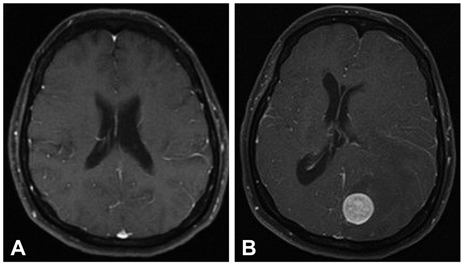
Fig. 1 A: Initial MRI performed in 2009 as part of staging for peripheral nerve sheath tumor. T1 weighted axial image post gadolinium injection was negative for abnormal intracranial enhancement. B: Repeat MRI performed August 2010 on admission to neurological intensive care unit. T1 weighted axial image post gadolinium injection revealed 2.2×2.2×2.2 cm, well-circumscribed, homogeneously enhancing lesion identified in the left occipital lobe with extensive vasogenic edema. A midline shift to the right of approximately 1 cm with subfalcine herniation is evident. Mass effect is identified on the occipital horn of the lateral ventricle.
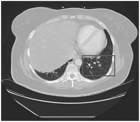
Fig. 2 Contrast-enhanced CT scan of the chest at the time of admission to the neurological intensive care unit. Scan revealed a new 1.1-cm left lower lobe solid nodular density adjacent to the lateral basilar segment artery and bronchus. A stable 0.4-cm nodule was seen peripheral to the left lower lobe.
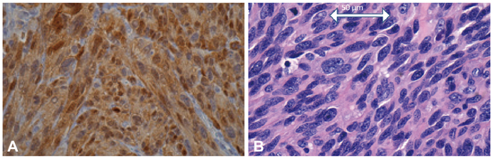
Fig. 3 A: Photomicrograph of an S-100 immunohistochemically stained slide (magnification ×400) of tissue from the original tumor, which confirms the tumor as an MPNST. B: Photomicrograph of an H & E slide (magnification ×400) of tissue from the cerebral mass. It shows a cellular neoplasm exhibiting storiform architecture with moderate nuclear atypia. Other areas show increased mitotic activity and necrosis. The diagnosis of MPNST was based on expression of similar immunohistochemical staining and nearly identical morphological characteristics shared between the original tumor and the metastatic tumor.
Reference
-
1. Fenzi F, Moretto G, Zamboni G, Passarin MG, Rizzuto N. Brain metastases from post-radiation malignant peripheral nerve sheath tumour. Ital J Neurol Sci. 1995. 16:495–498.
Article2. Tilgner J, Müller K, Ghanem N, Lutterbach J, Vesper J. Brain metastases as primary manifestation of a melanocytic malignant peripheral nerve sheath tumor in a 60-year-old man. BMC Neurol. 2007. 7:2.
Article3. McClatchey AI. Neurofibromatosis. Annu Rev Pathol. 2007. 2:191–216.
Article4. Kurokawa R, Tabuse M, Yoshida K, Kawase T. Spinal accessory schwannoma mimicking a tumor of the fourth ventricle: case report. Neurosurgery. 2004. 54:510–514. discussion 514.
Article5. Agrawal A, Rao KS, Makannavar JH, Shetty L, Raveendra VM. Intrasternomastoid spinal accessory nerve schwannoma: clinical and radiological correlation. Neurol India. 2005. 53:347–348.
Article6. McShane D, Noyek AM, Chapnik JS, Steinhardt MI, Cooter N. Schwannoma of the intrasternomastoid portion of the spinal accessory nerve: sophisticated preoperative CT diagnosis and appropriate surgical management. J Otolaryngol. 1986. 15:282–285.7. Noyek AM, Chapnik JS, Wortzman G, Kandel R. Schwannoma of the intrasternomastoid portion of the spinal accessory nerve: sophisticated pre-operative MRI diagnosis and appropriate surgical management. J Otolaryngol. 1992. 21:286–289.8. D'Agostino AN, Soule EH, Miller RH. Sarcomas of the peripheral nerves and somatic soft tissues associated with multiple neurofibromatosis (von recklinghausen's disease). Cancer. 1963. 16:1015–1027.9. Park SK, Yi HJ, Paik SS, Kim YJ, Ko Y, Oh SJ. Metastasizing malignant peripheral nerve sheath tumor initially presenting as intracerebral hemorrhage. Case report and review of the literature. Surg Neurol. 2007. 68:79–84. discussion 84.
Article10. Geller DS, Gebhardt M. Malignant peripheral nerve sheath tumors. Electronic Sarcoma Update Newsletter. 2006. Accessed 17 Feb 2011. Ossining, New York: Liddy Shriver Sarcoma Initiative;3. at http://sarcomahelp.org/learning_center/mpnst.html).11. Adamson DC, Cummings TJ, Friedman AH. Malignant peripheral nerve sheath tumor of the spine after radiation therapy for Hodgkin's lymphoma. Clin Neuropathol. 2004. 23:245–255.12. Amin A, Saifuddin A, Flanagan A, Patterson D, Lehovsky J. Radiotherapy-induced malignant peripheral nerve sheath tumor of the cauda equina. Spine (Phila Pa 1976). 2004. 29:E506–E509.
Article13. Friedrich RE, Kluwe L, Fünsterer C, Mautner VF. Malignant peripheral nerve sheath tumors (MPNST) in neurofibromatosis type 1 (NF1): diagnostic findings on magnetic resonance images and mutation analysis of the NF1 gene. Anticancer Res. 2005. 25:1699–1702.14. Poyhonen M, Niemela S, Herva R. Risk of malignancy and death in neurofibromatosis. Arch Pathol Lab Med. 1997. 121:139–143.15. Stojadinovic A, Yeh A, Brennan MF. Completely resected recurrent soft tissue sarcoma: primary anatomic site governs outcomes. J Am Coll Surg. 2002. 194:436–447.
Article16. Gupta G, Mammis A, Maniker A. Malignant peripheral nerve sheath tumors. Neurosurg Clin N Am. 2008. 19:533–543. v
Article
