J Clin Neurol.
2013 Jul;9(3):192-195. 10.3988/jcn.2013.9.3.192.
Transient Obstructive Hydrocephalus due to Intraventricular Hemorrhage: A Case Report and Review of Literature
- Affiliations
-
- 1Department of Neurological Surgery, Washington University School of Medicine, St. Louis, MO, USA. vellimana@wustl.edu
- 2Department of Neurosurgery, Swedish Medical Center, Seattle, WA, USA.
- KMID: 2287558
- DOI: http://doi.org/10.3988/jcn.2013.9.3.192
Abstract
- BACKGROUND
Acute transient obstructive hydrocephalus is rare in adults. We describe a patient with intraventricular hemorrhage (IVH) who experienced the delayed development of acute transient hydrocephalus.
CASE REPORT
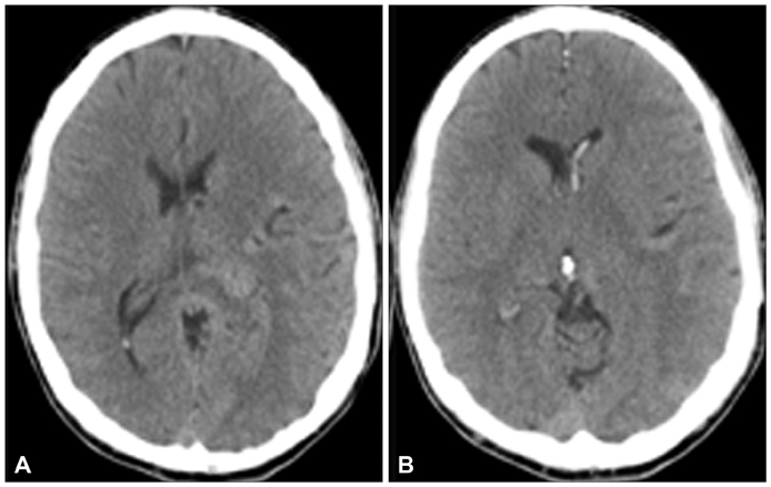
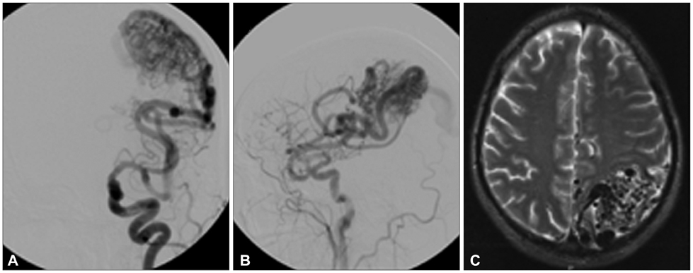
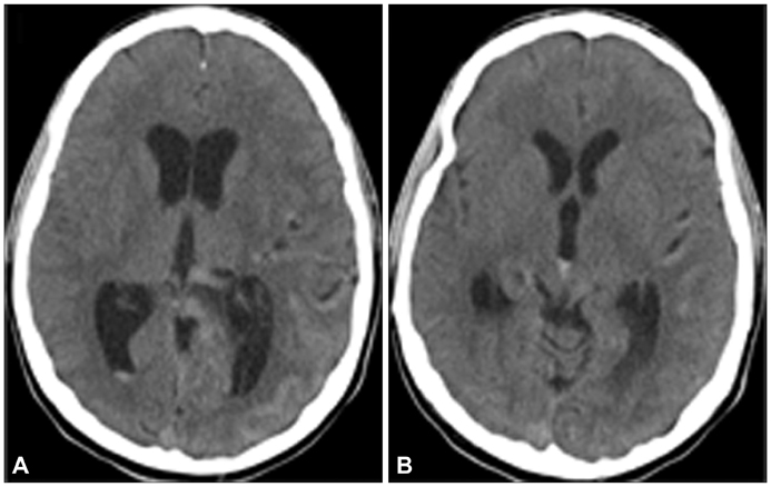
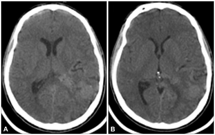
A 33-year-old man with a previously diagnosed Spetzler-Martin Grade 5 arteriovenous malformation presented with severe headache, which was found to be due to IVH. Forty hours after presentation he developed significant obstructive hydrocephalus due to the thrombus migrating to the cerebral aqueduct, and a ventriculostomy placement was planned. However, shortly thereafter his headache began to improve spontaneously. Within 4 hours after onset the headache had completely resolved, and an interval head CT scan revealed resolution of hydrocephalus.
CONCLUSIONS
In patients with IVH, acute obstructive hydrocephalus can develop at any time after the ictus. Though a delayed presentation of acute but transient obstructive hydrocephalus is unusual, it is important to be aware of this scenario and ensure that deterioration secondary to thrombus migration and subsequent obstructive hydrocephalus do not occur.
Keyword
MeSH Terms
Figure
Reference
-
1. Sharma RR, Chandy MJ, Lad SD. Transient hydrocephalus and acute lead encephalopathy in neonates and infants. Report of two cases. Br J Neurosurg. 1990; 4:141–145.
Article2. Prabhu SS, Sharma RR, Gurusinghe NT, Parekh HC. Acute transient hydrocephalus in carbon monoxide poisoning: a case report. J Neurol Neurosurg Psychiatry. 1993; 56:567–568.
Article3. Dubey AK, Rao KL. Pathology of post meningitic hydrocephalus. Indian J Pediatr. 1997; 64:6 Suppl. 30–33.
Article4. Abubacker M, Bosma JJ, Mallucci CL, May PL. Spontaneous resolution of acute obstructive hydrocephalus in the neonate. Childs Nerv Syst. 2001; 17:182–184.
Article5. Hagihara N, Abe T, Inoue K, Watanabe M, Tabuchi K. Rapid resolution of hydrocephalus due to simultaneous movements of hematoma in the trigono-occipital horn and the aqueduct. Neurol India. 2009; 57:357–358.
Article6. Inamura T, Kawamura T, Inoha S, Nakamizo A, Fukui M. Resolving obstructive hydrocephalus from AVM. J Clin Neurosci. 2001; 8:569–570.
Article7. Yoshimoto Y, Ochiai C, Kawamata K, Endo M, Nagai M. Aqueductal blood clot as a cause of acute hydrocephalus in subarachnoid hemorrhage. AJNR Am J Neuroradiol. 1996; 17:1183–1186.8. Gupta SK, Sharma T. Acute post-traumatic hydrocephalus in an infant due to aqueductal obstruction by a blood clot: a case report. Childs Nerv Syst. 2009; 25:373–376.
Article9. Khan R, Mamourian AC, Radwan T. Utility of multislice computed tomography and reformatted images: identification of migratory intraventricular clot exacerbating obstructive hydrocephalus. J Neurosurg. 2008; 109:156–158.
Article10. Tsementzis SA, Honan WP, Nightingale S, Hitchcock ER, Meyer CH. Fibrinolytic activity after subarachnoid haemorrhage and the effect of tranexamic acid. Acta Neurochir (Wien). 1990; 103:116–121.
Article11. Wu KK, Jacobsen CD, Hoak JC. Plasminogen in normal and abnormal human cerebrospinal fluid. Arch Neurol. 1973; 28:64–66.
Article12. Tovi D, Nilsson IM, Thulin CA. Fibrinolytic activity of the cerebrospinal fluid after subarachnoid haemorrhage. Acta Neurol Scand. 1973; 49:1–9.
Article13. Quencer RM. Intracranial CSF flow in pediatric hydrocephalus: evaluation with cine-MR imaging. AJNR Am J Neuroradiol. 1992; 13:601–608.14. Nomura S, Orita T, Tsurutani T, Kajiwara K, Izumihara A. Transient hydrocephalus due to movement of a clot plugging the aqueduct. Comput Med Imaging Graph. 1997; 21:351–353.
Article
- Full Text Links
- Actions
-
Cited
- CITED
-
- Close
- Share
- Similar articles
-
- A Case of congenital hydrocephalus associated with fetal intraventricular hemorrhage
- Intraventricular Glioblastoma Multiforme with Previous History of Intracerebral Hemorrhage : A Case Report
- Traumatic Intraventricular Hemorrhage: 2 Cases Report
- Endoscopic surgery for obstructive hydrocephalus
- Fibrinolytic (Thrombolytic) Therapy for Post Intraventricular Hemorrhagic Hydrocephalus in Preterm Infants


