J Breast Cancer.
2010 Jun;13(2):227-230. 10.4048/jbc.2010.13.2.227.
Ectopic Extramammary Paget's Disease of the Breast Skin: A Case Report
- Affiliations
-
- 1Department of Surgery, Seoul National University Boramae Hospital, Seoul, Korea.
- 2Department of Pathology, Seoul National University Boramae Hospital, Seoul, Korea.
- 3Department of Dermatology, Seoul National University Boramae Hospital, Seoul, Korea.
- 4Cancer Research Institute and Department of Surgery, College of Medicine, Seoul National University, Seoul, Korea. dynoh@plaza.snu.ac.kr
- KMID: 2286550
- DOI: http://doi.org/10.4048/jbc.2010.13.2.227
Abstract
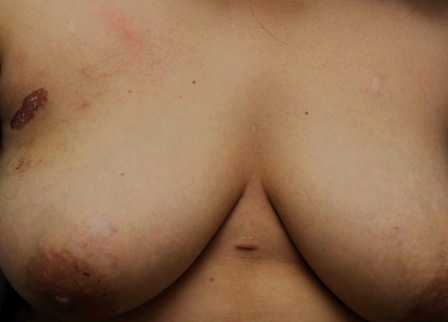
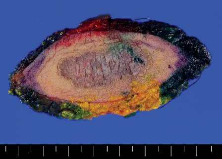
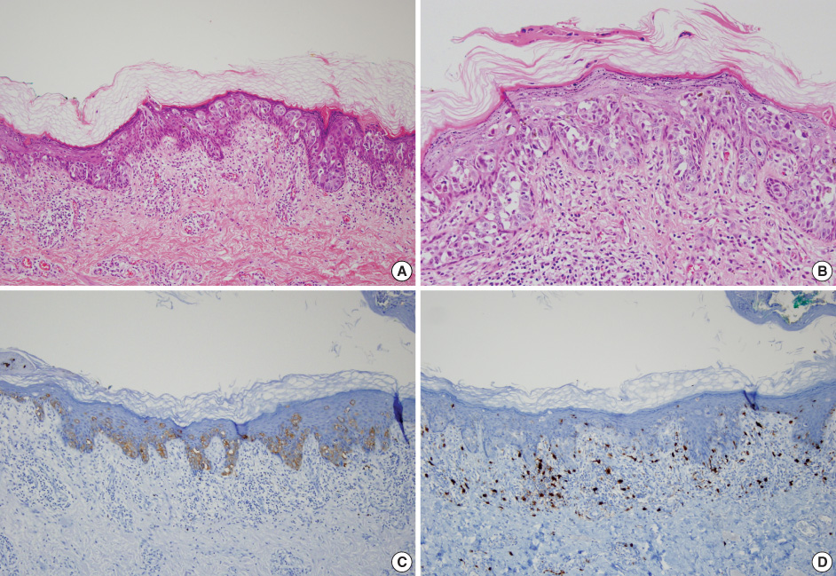
- Whereas extramammary Paget's disease commonly occurs in the apocrine gland rich skin areas, ectopic extramammary Paget's disease develops in the skin areas that are devoid of apocrine glands. We experienced the case of a 34 year-old female patient who had a skin lesion in the upper outer quadrant of the right breast for 5 years and that lesion was diagnosed as Paget's disease according to the punch biopsy. There was no other underlying malignancy, and so wide excision was performed. The final pathologic diagnosis was Paget's disease confined to the epidermis and the size of the tumor was 3.0x1.1 cm. Positive staining for cytokeratin 7, epithelial membrane antigen and negative staining for S-100 protein and HMB-45 was observed on the immunohistochemical tests. We report here on an extremely unusual case of ectopic extramammary Paget's disease of the breast skin, and we include a review of the relevant literature.
Keyword
MeSH Terms
Figure
Reference
-
1. Paget J. On disease of the mammary areola preceding cancer of the mammary gland. St Bartholomew Hosp Res Lond. 1874. 10:87–89. Cited from Kanitakis J. Mammary and extramammary Paget's disease. J Eur Acad Dermatol Venereol 2007;21:581-90.
Article2. Ashikari R, Park K, Huvos AG, Urban JA. Paget's disease of the breast. Cancer. 1970. 26:680–685.
Article3. Fardal RW, Kierland RR, Clagett OT, Woolner LB. Prognosis in cutaneous Paget's disease. Postgrad Med. 1964. 36:584–593.
Article4. Crocker H. Paget's disease affecting the scrotum and penis. Trans Pathol Soc London. 1889. 40:187–191. Cited from Kanitakis J. Mammary and extramammary Paget's disease. J Eur Acad Dermatol Venereol 2007;21:581-90.5. Youn HJ, Jung SH, Kim JC. A case of extramammary Paget's disease of the axilla. J Breast Cancer. 2008. 11:156–159.
Article6. Kanitakis J. Mammary and extramammary Paget's disease. J Eur Acad Dermatol Venereol. 2007. 21:581–590.
Article7. Cohen MA, Hanly A, Poulos E, Goldstein GD. Extramammary Paget's disease presenting on the face. Dermatol Surg. 2004. 30:1361–1363.
Article8. McCarter MD, Quan SH, Busam K, Paty PP, Wong D, Guillem JG. Long-term outcome of perianal Paget's disease. Dis Colon Rectum. 2003. 46:612–616.
Article9. Salamanca J, Benito A, García-Peñalver C, Azorín D, Ballestín C, Rodríguez-Peralto JL. Paget's disease of the glans penis secondary to transitional cell carcinoma of the bladder: a report of two cases and review of the literature. J Cutan Pathol. 2004. 31:341–345.
Article10. Inoue S, Aki T, Mihara M. Paget's disease in siblings. Dermatology. 2000. 201:178.11. Lloyd J, Flanagan AM. Mammary and extramammary Paget's disease. J Clin Pathol. 2000. 53:742–749.
Article12. Murata Y, Kumano K. Extramammary Paget's disease of the genitalia with clinically clear margins can be adequately resected with 1 cm margin. Eur J Dermatol. 2005. 15:168–170.13. Brash DE. De Vita VT, Hellman S, Rosenberg SA, editors. Cancers of the skin. Cancer: Principles and practice of Oncology. 1997. 5th ed. New York: Lippincott Raven;1565–1566.14. Hendi A, Brodland DG, Zitelli JA. Extramammary Paget's disease: surgical treatment with Mohs micrographic surgery. J Am Acad Dermatol. 2004. 51:767–773.
Article15. Hatta N, Morita R, Yamada M, Echigo T, Hirano T, Takehara K, et al. Sentinel lymph node biopsy in patients with extramammary Paget's disease. Dermatol Surg. 2004. 30:1329–1334.
Article16. Chanda JJ. Extramammary Paget's disease: prognosis and relationship to internal malignancy. J Am Acad Dermatol. 1985. 13:1009–1014.
Article


