J Breast Cancer.
2010 Dec;13(4):349-356. 10.4048/jbc.2010.13.4.349.
Evaluation of Phase-Contrast Microscopic Imaging with Synchrotron Radiation in the Diagnosis of Breast Cancer and Differentiation of Various Breast Diseases: Preliminary Results
- Affiliations
-
- 1Department of Surgery, Catholic University of Daegu School of Medicine, Daegu, Korea. shwpark@cu.ac.kr
- 2Department of Anatomy, Catholic University of Daegu School of Medicine, Daegu, Korea.
- 3Department of Radiology and Biomedical Engineering, Catholic University of Daegu School of Medicine, Daegu, Korea.
- 4Department of Pathology, Catholic University of Daegu School of Medicine, Daegu, Korea.
- 5Pohang Accelerator Laboratory, Pohang University of Science and Technology, Pohang, Korea.
- KMID: 2286522
- DOI: http://doi.org/10.4048/jbc.2010.13.4.349
Abstract
- PURPOSE
A significant improvement of imaging using synchrotron radiation (SR) is obtained by introducing phase-contrast technique. This technique provides greatly enhanced contrast and good soft tissue discrimination with high spatial resolution. The aim of this study was to observe microstructures of pathologic breast specimens including invasive breast cancer using phase-contrast technique with SR and to evaluate the feasibility of phase-contrast imaging in clinical application.
METHODS
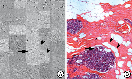
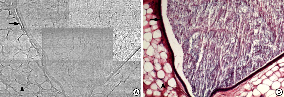
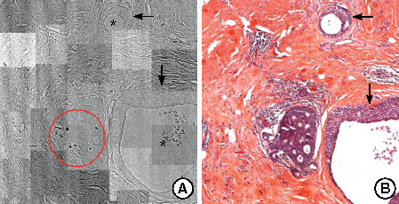
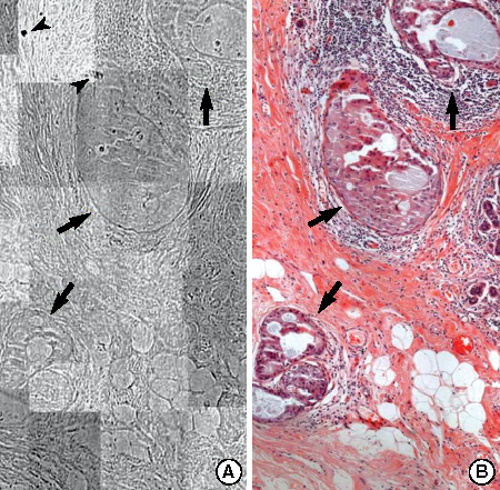
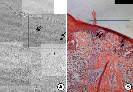
Phase-contrast microscopic image of normal breast tissue and the images of various breast diseases such as fibrocystic change, ductal carcinoma in situ, invasive ductal carcinoma, Paget's disease were obtained using hard X-ray microscopy with an 11.1 keV monochromatic beam from SR source and CsI (TI) scintillation crystal. Zernike phase-shifter was adapted for phase-contrast hard X-ray microscopy. The visual image was magnified 20 times by microscopic objective lens and captured using a full frame charge-coupled device camera. Obtained images were compared with corresponding histopathologic findings in the optical microscopy.
RESULTS
The SR images of various breast diseases were obtained with a good contrast and high visibility by phase-contrast technique. It was possible to observe the microstructures with high spatial resolution down to the micron region. The characteristic features of each disease were consistent with the histopathologic findings of corresponding sample and the images of breast cancer and the other diseases were distinct from each other.
CONCLUSION
Using phase-contrast technique, SR images of various breast diseases including breast cancer were obtained. These images were comparable with standard histopathologic findings and showed different features for each disease. The results suggest that phase-contrast microscopic imaging with SR has potential as a diagnostic tool and also its clinical application is feasible, especially in breast imaging.
MeSH Terms
Figure
Reference
-
1. Housa D, Housová J, Vernerová Z, Haluzik M. Adipocytokines and cancer. Physiol Res. 2006. 55:233–244.
Article2. Jatoi I, Miller AB. Why is breast cancer mortality declining? Lancet Oncol. 2003. 4:251–254.3. Margaritondo G, Meuli R. Synchrotron radiation in radiology: novel X-ray sources. Eur Radiol. 2003. 13:2633–2641.
Article4. Lewis RA, Rogers KD, Hall CJ, Hufton AP, Evans S, Menk RH, et al. Antonuk LE, Yaffe MJ, editors. Diffraction enhanced imaging: improved contrast, lower dose Xray imaging. Medical Imaging 2002: Physics of Medical Imaging, San Diego, USA. 2002. Bellingham: Society of Photo-optical Instrumentation Engineers;286–297.5. Lewis RA. Meidcal phase contrast X-ray imaging: current status and future prospects. Phys Med Biol. 2004. 49:3573–3583.6. Suortti P, Thomlinson W. Medical applications of synchrotron radiation. Phys Med Biol. 2003. 48:R1–R35.
Article7. Burattini E, Cossu E, Maggio C, Gambaccini M, Indovina PL, Marziani M, et al. Mammography with synchrotron radiation. Radiology. 1995. 195:239–244.
Article8. Arfelli F, Bonvicini V, Bravin A, Cantatore G, Castelli E, Palma LD, et al. Mammography with synchrotron radiation: phase-detection techniques. Radiology. 2000. 215:286–293.
Article9. Ingal VN, Beliaevskaya EA, Brianskaya AP, Merkurieva RD. Phase mammography: a new technique for breast investigation. Phys Med Biol. 1998. 43:2555–2567.10. Liu C, Yan X, Zhang X, Yang W, Peng W, Shi D, et al. Evaluation of X-ray diffraction enhanced imaging in the diagnosis of breast cancer. Phys Med Biol. 2007. 52:419–427.
Article11. Fiedler S, Bravin A, Keyriläinen J, Fernández M, Suortti P, Thomlinson W, et al. Imaging lobular breast carcinoma: comparison of synchrotron radiation DEI-CT technique with clinical CT, mammography and histology. Phys Med Biol. 2004. 49:175–188.
Article12. Jeong YJ, Bong JG, Kim HT, Kim JK, Jheon SH, Youn HS, et al. Synchrotron radiation imaging of female breast tissues using phase contrast technique. J Breast Cancer. 2008. 11:40–44.
Article13. Park SH, Kim HT, Kim JK, Jheon SH, Youn HS. Hard X-ray microscopic imaging of human breast tissues. 2006. In : 9th International Conference on Synchrotron Radiation Instrumentation; Daegu: American Institute of Physics.14. Stuart JS, Anthony JG. Harris JR, Lippman ME, Morrow M, Osborne CK, editors. Pathology of invasive breast cancer. Diseases of the Breast. 2004. 3rd ed. Philadelphia: Lippincott Williams & Wilkins;542–584.15. Azzopardi JG. Azzopardi JG, Ahmed A, Millis RR, editors. Epitheliosis and in situ carcinoma. Problems in Breast Pathology. 1979. Philadelphia: Saunders;128.16. Rosen PP. Rosen PP, editor. Paget's disease of the nipple. Rosen' Breast Pathology. 1997. Philadelphia: Lippincott-Raven;493–506.17. Youn HS, Jung SW. Hard X-ray microscopy with Zernike phase contrast. J Microsc. 2006. 223(Pt 1):53–56.
Article18. Weon BM, Je JH, Hwu Y, Margaritondo G. Phase contrast X-ray imaging. Int J Nanotechnol. 2006. 3:280–297.
Article19. Socha JJ, Westneat MW, Harrison JF, Waters JS, Lee WK. Real-time phase-contrast X-ray imaging: a new technique for the study of animal form and function. BMC Biol. 2007. 5:6.
Article20. Kagoshima Y, Yokoyama Y, Niimi T, Koyama T, Tsusaka Y, Matsui J, et al. Hard X-ray phase-contrast microscope for observing transparent specimens. 2002. In : 7th International Conference on X-Ray Microscopy; Grenoble: European Synchrotron Radiation Facility.21. Andrews JC, Almeida E, van der Meulen MC, Alwood JS, Lee C, Liu Y, et al. Nanoscale X-ray microscopic imaging of mammalian mineralized tissue. Microsc Microanal. 2010. 16:327–336.
Article22. Chen J, Yang Y, Zhang X, Antrews JC, Pianetta P, Guan Y, et al. 3D nanoscale imaging of the yeast, Schizosaccharomyces pombe, by full-field transmission X-ray microscopy at 5.4 keV. Anal Bioanal Chem. 2010. 397:2117–2121.
Article23. Youn HS, Baik SY, Chang CH. Hard X-ray microscopy with a 130 nm spatial resolution. Rev Sci Instrum. 2005. 76:023702.24. Schiffhauer LM, Boger JN, Bonfiglio TA, Zavislan JM, Zuley M, Fox CA. Confocal microscopy of unfixed breast needle core biopsies: a comparison to fixed and stained sections. BMC Cancer. 2009. 9:265.
Article25. Huang D, Swanson EA, Lin CP, Schuman JS, Stinson WG, Chang W, et al. Optical coherence tomography. Science. 1991. 254:1178–1181.
Article26. Fercher AF, Drexler W, Hitzenberger CK, Lasser T. Optical coherence tomography: principles and applications. Rep Prog Phys. 2003. 66:239–303.27. Pani S, Longo R, Derossi D, Montanari F, Olivo A, Arfelli F, et al. Breast tomography with synchrotron radiation: preliminary results. Phys Med Biol. 2004. 49:1739–1754.
Article28. Lindfors KK, Boone JM, Nelson TR, Yang K, Kwan AL, Miller DF. Dedicated breast CT: initial clinical experience. Radiology. 2008. 246:725–733.
Article29. Arfelli F, Assante M, Bonvicini V, Bravin A, Cantatore G, Castelli E, et al. Low-dose phase contrast X-ray medical imaging. Phys Med Biol. 1998. 43:2845–2852.
Article30. Boone JM, Nelson TR, Lindfors KK, Seibert JA. Dedicated breast CT: radiation dose and image quality evaluation. Radiology. 2001. 221:657–667.
Article
- Full Text Links
- Actions
-
Cited
- CITED
-
- Close
- Share
- Similar articles
-
- Synchrotron Radiation Imaging of Breast Tissue Using a Phase-contrast Hard X-ray Microscope
- Synchrotron Radiation Imaging of Female Breast Tissues Using Phase Contrast Technique
- Magnetic Resonance Findings of Breast Diseases
- 3D Histology Using the Synchrotron Radiation Propagation Phase Contrast Cryo-microCT
- Optical Imaging of the Breast


