The Effects of Preoperative 18F-FDG PET/CT in Breast Cancer Patients in Comparison to the Conventional Imaging Study
- Affiliations
-
- 1Department of Surgery, Eulji University Hospital, Eulji University School of Medicine, Daejeon, Korea. yj0139@naver.com
- 2Department of Anesthesiology, Chungbuk National University Hospital, Chungbuk National University School of Medicine, Cheongju, Korea.
- 3Department of Nuclear Medicine, Eulji University Hospital, Eulji University School of Medicine, Daejeon, Korea.
- KMID: 2286442
- DOI: http://doi.org/10.4048/jbc.2012.15.4.441
Abstract
- PURPOSE
There have been recent studies of the 18F-fluorodeoxyglucose positron emission tomography and computed tomography (18F-FDG PET/CT) in the staging, detection, and follow-up of the breast cancer occurrence and recurrence. There was controversy concerning the use of 18F-FDG PET/CT for staging primary breast cancer. In this study, we investigated the potential effects of 18F-FDG PET/CT in the initial assessment of patients with primary breast cancer.
METHODS
From January 2008 to December 2009, 154 consecutive biopsy-proven invasive breast cancer patients were enrolled in this study. Patients underwent conventional imaging studies including mammography, breast ultrasonography (USG), and magnetic resonance imaging for local assessment, and plain chest X-ray, liver USG, and bone scan to rule out distant metastasis. All 154 patients underwent 18F-FDG PET/CT in the initial assessment.
RESULTS
18F-FDG PET/CT did not detect primary breast lesions in 16 patients with a sensitivity of 89.6% and detected only 5 multiple lesions (12.5%) out of 40 cases. Histologically confirmed axillary lymph node (LN) metastases were in 51 patients, and the sensitivity and specificity of 18F-FDG PET/CT to detect metastatic axilla were 37.3% and 95.8%, respectively; whereas the corresponding estimates of USG were 41.2% and 93.7%, respectively. Eleven extra-axillary LN metastases were found in eight patients, and seven lesions were detected by 18F-FDG PET/CT only. The sensitivity and specificity of 18F-FDG PET/CT in detecting distant metastasis were 100% and 96.4%, respectively; whereas the sensitivity and specificity of the conventional imaging were 61.5% and 99.2%, respectively.
CONCLUSION
18F-FDG PET/CT cannot be recommended as a primary diagnostic procedure in breast cancer, but it has the potential to be used as an additional imaging tool for the detection of axillary metastasis, distant metastasis, and extra-axillary LN metastasis. 18F-FDG PET/CT cannot solely replace the conventional diagnostic procedure in primary breast cancer. The best approach may be the combination of different imaging modalities.
MeSH Terms
-
Axilla
Breast
Breast Neoplasms
Diagnostic Imaging
Fluorodeoxyglucose F18
Follow-Up Studies
Humans
Liver
Lymph Nodes
Magnetic Resonance Imaging
Mammography
Neoplasm Metastasis
Positron-Emission Tomography
Positron-Emission Tomography and Computed Tomography
Recurrence
Sensitivity and Specificity
Thorax
Ultrasonography, Mammary
Fluorodeoxyglucose F18
Figure
-
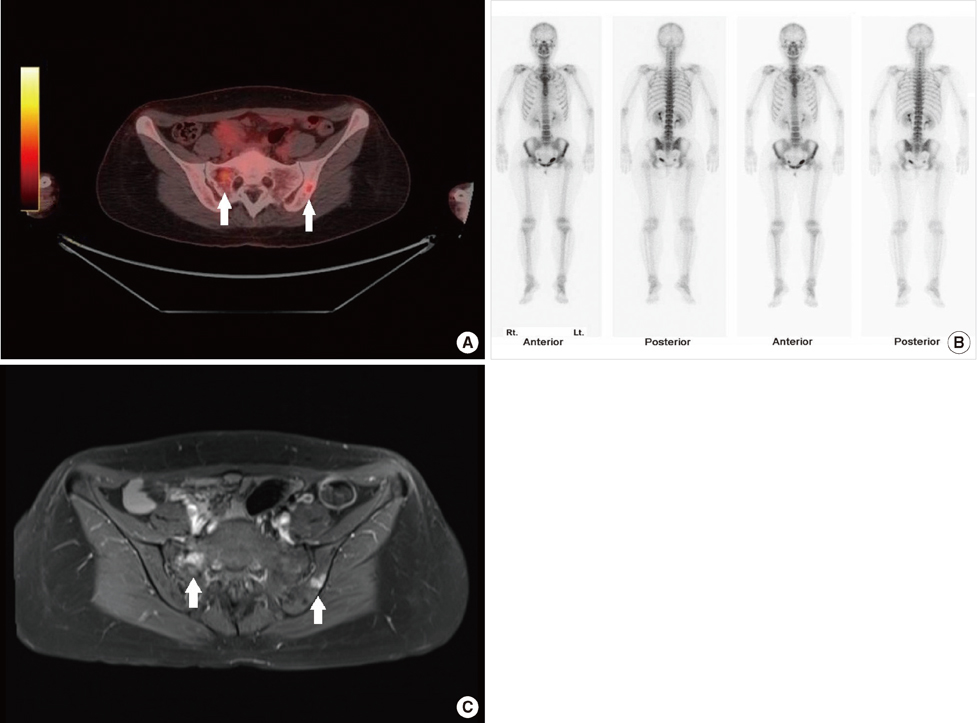
Figure 1 Image findings of a 38-year-old breast cancer patient with multiple bone metastases. Mild hypermetabolism were seen at right sacrum and left iliac bone on 18F-fluorodeoxyglucose positron emission tomography and computed tomography (A, arrows) which was equivocal on bone scan (B). The lesions revealed as osteolytic bone metastasis with low SI on T1- and increased SI on T2 weighted images, with strong enhancement on contrast enhanced magnetic resonance imaging (C, arrows).
Cited by 2 articles
-
Polyostotic Fibrous Dysplasia Mimicking Multiple Bone Metastases in a Patient with Ductal Carcinoma In Situ
Jun Ho Lee, Sung Yong Kim, Jeong Eon Lee, Eun Yoon Cho, Yoon-La Choi, Joon Young Choi, Sun Wook Han, Seok Won Kim, Won Ho Kil, Seok Jin Nam
J Breast Cancer. 2014;17(1):83-87. doi: 10.4048/jbc.2014.17.1.83.Effectiveness of Breast MRI and 18F-FDG PET/CT for the Preoperative Staging of Invasive Lobular Carcinoma versus Ductal Carcinoma
Na Young Jung, Sung Hoon Kim, Sung Hun Kim, Ye Young Seo, Jin Kyoung Oh, Hyun Su Choi, Won Jong You
J Breast Cancer. 2015;18(1):63-72. doi: 10.4048/jbc.2015.18.1.63.
Reference
-
1. Eubank WB, Mankoff DA, Takasugi J, Vesselle H, Eary JF, Shanley TJ, et al. 18fluorodeoxyglucose positron emission tomography to detect mediastinal or internal mammary metastases in breast cancer. J Clin Oncol. 2001. 19:3516–3523.
Article2. Fehr MK, Hornung R, Varga Z, Burger D, Hess T, Haller U, et al. Axillary staging using positron emission tomography in breast cancer patients qualifying for sentinel lymph node biopsy. Breast J. 2004. 10:89–93.
Article3. Jacobs MA, Ouwerkerk R, Wolff AC, Gabrielson E, Warzecha H, Jeter S, et al. Monitoring of neoadjuvant chemotherapy using multiparametric, 23Na sodium MR, and multimodality (PET/CT/MRI) imaging in locally advanced breast cancer. Breast Cancer Res Treat. 2011. 128:119–126.
Article4. Pennant M, Takwoingi Y, Pennant L, Davenport C, Fry-Smith A, Eisinga A, et al. A systematic review of positron emission tomography (PET) and positron emission tomography/computed tomography (PET/CT) for the diagnosis of breast cancer recurrence. Health Technol Assess. 2010. 14:1–103.
Article5. Kumar R, Zhuang H, Schnall M, Conant E, Damia S, Weinstein S, et al. FDG PET positive lymph nodes are highly predictive of metastasis in breast cancer. Nucl Med Commun. 2006. 27:231–236.
Article6. Zornoza G, Garcia-Velloso MJ, Sola J, Regueira FM, Pina L, Beorlegui C. 18F-FDG PET complemented with sentinel lymph node biopsy in the detection of axillary involvement in breast cancer. Eur J Surg Oncol. 2004. 30:15–19.
Article7. Yang SN, Liang JA, Lin FJ, Kao CH, Lin CC, Lee CC. Comparing whole body (18)F-2-deoxyglucose positron emission tomography and technetium-99m methylene diphosphonate bone scan to detect bone metastases in patients with breast cancer. J Cancer Res Clin Oncol. 2002. 128:325–328.8. Ueda S, Saeki T, Shigekawa T, Omata J, Moriya T, Yamamoto J, et al. 18F-fluorodeoxyglucose positron emission tomography optimizes neoadjuvant chemotherapy for primary breast cancer to achieve pathological complete response. Int J Clin Oncol. 2012. 17:276–282.
Article9. Segaert I, Mottaghy F, Ceyssens S, De Wever W, Stroobants S, Van Ongeval C, et al. Additional value of PET-CT in staging of clinical stage IIB and III breast cancer. Breast J. 2010. 16:617–624.
Article10. Inokuchi M, Furukawa H, Kayahara M, Ohta T, Kawashima H, Taki J, et al. The role of / 18FDG PET/CT for the initial staging and therapy in primary breast cancer. Gan To Kagaku Ryoho. 2009. 36:2526–2531.11. Bombardieri E, Crippa F. PET imaging in breast cancer. Q J Nucl Med. 2001. 45:245–256.12. Garami Z, Hascsi Z, Varga J, Dinya T, Tanyi M, Garai I, et al. The value of 18-FDG PET/CT in early-stage breast cancer compared to traditional diagnostic modalities with an emphasis on changes in disease stage designation and treatment plan. Eur J Surg Oncol. 2012. 38:31–37.
Article13. Danforth DN Jr, Aloj L, Carrasquillo JA, Bacharach SL, Chow C, Zujewski J, et al. The role of 18F-FDG-PET in the local/regional evaluation of women with breast cancer. Breast Cancer Res Treat. 2002. 75:135–146.
Article14. Veronesi U, De Cicco C, Galimberti VE, Fernandez JR, Rotmensz N, Viale G, et al. A comparative study on the value of FDG-PET and sentinel node biopsy to identify occult axillary metastases. Ann Oncol. 2007. 18:473–478.
Article15. Robertson IJ, Hand F, Kell MR. FDG-PET/CT in the staging of local/regional metastases in breast cancer. Breast. 2011. 20:491–494.
Article16. Heusner TA, Freudenberg LS, Kuehl H, Hauth EA, Veit-Haibach P, Forsting M, et al. Whole-body PET/CT-mammography for staging breast cancer: initial results. Br J Radiol. 2008. 81:743–748.
Article17. Bernsdorf M, Berthelsen AK, Wielenga VT, Kroman N, Teilum D, Binderup T, et al. Preoperative PET/CT in early-stage breast cancer. Ann Oncol. 2012. 23:2277–2282.
Article18. Fuster D, Duch J, Paredes P, Velasco M, Muñoz M, Santamaria G, et al. Preoperative staging of large primary breast cancer with [18F]fluorodeoxyglucose positron emission tomography/computed tomography compared with conventional imaging procedures. J Clin Oncol. 2008. 26:4746–4751.
Article19. Lerman H, Metser U, Grisaru D, Fishman A, Lievshitz G, Even-Sapir E. Normal and abnormal 18F-FDG endometrial and ovarian uptake in pre- and postmenopausal patients: assessment by PET/CT. J Nucl Med. 2004. 45:266–271.20. Liu Y. Benign ovarian and endometrial uptake on FDG PET-CT: patterns and pitfalls. Ann Nucl Med. 2009. 23:107–112.
Article21. Alkhawaldeh K, Bural G, Kumar R, Alavi A. Impact of dual-time-point (18)F-FDG PET imaging and partial volume correction in the assessment of solitary pulmonary nodules. Eur J Nucl Med Mol Imaging. 2008. 35:246–252.
Article22. Kumar R, Halanaik D, Malhotra A. Clinical applications of positron emission tomography-computed tomography in oncology. Indian J Cancer. 2010. 47:100–119.
Article23. Ohta M, Tokuda Y, Suzuki Y, Kubota M, Makuuchi H, Tajima T, et al. Whole body PET for the evaluation of bony metastases in patients with breast cancer: comparison with 99Tcm-MDP bone scintigraphy. Nucl Med Commun. 2001. 22:875–879.
Article
- Full Text Links
-
- Actions
-
Cited
- CITED
-
- Close
- Share
-
- Similar articles
-
- Use of 18F-FDG PET/CT in Second Primary Cancer
- 18F-FDG PET/CT Findings in a Breast Cancer Patient with Concomitant Tuberculous Axillary Lymphadenitis
- Preoperative Axillary Staging Using 18F-FDG PET/CT and Ultrasonography in Breast Cancer Patients
- Clinical Significance of Focal Breast Lesions Incidentally Identified by 18F-FDG PET/CT
- Comparison of CT or MRI and 18F-FDG PET/CT for the Preoperative Staging Accuracy of Ovarian Cancer
