Diagnostic Value of Contrast-Enhanced Ultrasound Parametric Imaging in Breast Tumors
- Affiliations
-
- 1Department of Ultrasound, Pudong New Area People's Hospital, Shanghai, China. doct1167@126.com
- KMID: 2286403
- DOI: http://doi.org/10.4048/jbc.2013.16.2.208
Abstract
- PURPOSE
This study aimed to evaluate the diagnostic value of SonoLiver software for parametric imaging in breast tumors.
METHODS
Contrast-enhanced ultrasound (CEUS) was performed in 216 breast lesions (113 malignant, 103 benign). The CEUS parameters were compared between benign and malignant lesions. The rise time, the time to peak, the mean transit time and dynamic vessel pattern (DVP) were analyzed using SonoLiver software.
RESULTS
Quantitative analysis showed that the rise time was 16.52+/-4.15 seconds in the benign group vs. 13.86+/-3.36 seconds in the malignant group (p=0.007), and the time to peak was 19.86+/-4.87 seconds in the benign group vs. 16.52+/-4.85 seconds in the malignant group (p=0.009). The mean transit time was 80.55+/-18.65 seconds in the benign group vs. 65.16+/-20.28 seconds in the malignant group (p=0.006). The difference between the distribution of DVP in benign and malignant tumors was statistically significant. One hundred one malignant tumors (89.4%) performed an irregular red/yellow fill in the region of interest (ROI) and 85 benign tumors (82.5%) performed a single blue/green fill in the ROI. The sensitivity, specificity, and accuracy of parametric imaging in breast tumors were 84.1%, 85.4%, 84.7%, respectively.
CONCLUSION
The CEUS parametric imaging can distinguish differences between malignant and benign breast tumors as well as provide diagnostic information on breast lesions.
Keyword
MeSH Terms
Figure
-
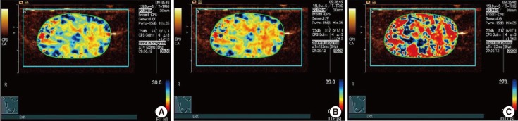
Figure 1 In a 59-year-old woman with a palpable mass in the inner upper quadrant of her left breast, surgical pathology proved an invasive ductal carcinoma. Rise time (RT) parametric imaging was given 1 point (A), time to peak (TTP) parametric imaging was given 1 point (B), and mean transit time (mTT) parametric imaging was given 1 point (C). Finally, the total score of this case was 3 (1+1+1=3).
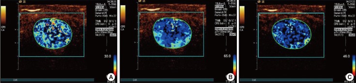
Figure 2 In a 35-year-old woman with a palpable mass in the outer upper quadrant of her right breast, surgical pathology proved a fibroadenoma. Rise time (RT) parametric imaging was given 0 points (A), time to peak (TTP) parametric imaging was given 0 points (B), and mean transit time (mTT) parametric imaging was given 0 points (C). Finally, the total score of this case was 0 (0+0+0=0).
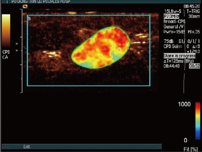
Figure 3 In a 68-year-old woman with a palpable mass in the outer upper quadrant of her left breast, surgical pathology proved an invasive ductal carcinoma. Distribution of dynamic vessel pattern performed a red/yellow fill.
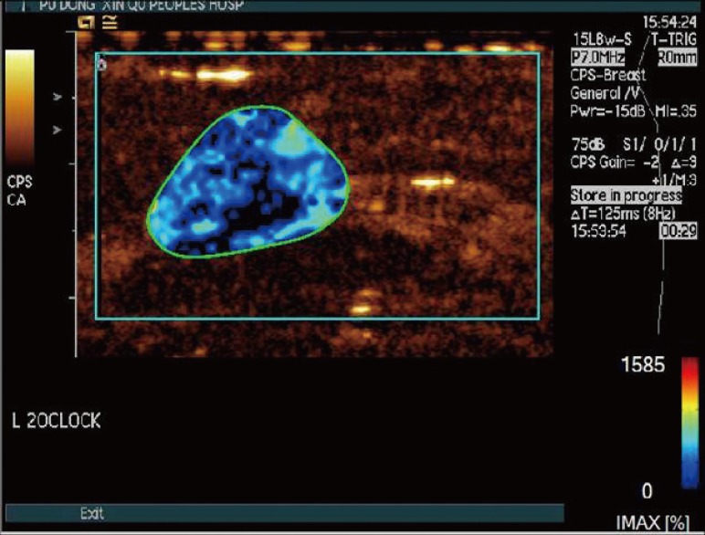
Figure 4 In a 25-year-old woman with a palpable mass in the inner upper quadrant of her left breast, surgical pathology proved a fibroadenoma. Distribution of dynamic vessel pattern performed a single blue/green fill.

Figure 5 In a 31-year-old woman with a palpable mass in the inner upper quadrant of her right breast, surgical pathology proved a fibroadenoma. Distribution of dynamic vessel pattern performed a few points of linear red/yellow fill and most of the lesion were a blue/green fill.
Reference
-
1. Zhao H, Xu R, Ouyang Q, Chen L, Dong B, Huihua Y. Contrast-enhanced ultrasound is helpful in the differentiation of malignant and benign breast lesions. Eur J Radiol. 2010; 73:288–293. PMID: 19559551.
Article2. Ricci P, Cantisani V, Ballesio L, Pagliara E, Sallusti E, Drudi FM, et al. Benign and malignant breast lesions: efficacy of real time contrast-enhanced ultrasound vs. magnetic resonance imaging. Ultraschall Med. 2007; 28:57–62. PMID: 17304413.
Article3. Du J, Li FH, Fang H, Xia JG, Zhu CX. Microvascular architecture of breast lesions: evaluation with contrast-enhanced ultrasonographic micro flow imaging. J Ultrasound Med. 2008; 27:833–842. PMID: 18499843.4. Jiang YX, Liu H, Liu JB, Zhu QL, Sun Q, Chang XY. Breast tumor size assessment: comparison of conventional ultrasound and contrast-enhanced ultrasound. Ultrasound Med Biol. 2007; 33:1873–1881. PMID: 17686569.
Article5. Saracco A, Aspelin P, Leifland K, Themudo R, Wilczek B, Axelsson R. Bolus compared with continuous infusion of microbubble contrast agent using real-time contrast harmonic imaging ultrasound in breast tumors. Acta Radiol. 2009; 50:854–859. PMID: 19634024.
Article6. Sorelli PG, Cosgrove DO, Svensson WE, Zaman N, Satchithananda K, Barrett NK, et al. Can contrast-enhanced sonography distinguish benign from malignant breast masses? J Clin Ultrasound. 2010; 38:177–181. PMID: 20146214.
Article7. Barnard S, Leen E, Cooke T, Angerson W. A contrast-enhanced ultrasound study of benign and malignant breast tissue. S Afr Med J. 2008; 98:386–391. PMID: 18637311.8. Kettenbach J, Helbich TH, Huber S, Zuna I, Dock W. Computer-assisted quantitative assessment of power Doppler US: effects of microbubble contrast agent in the differentiation of breast tumors. Eur J Radiol. 2005; 53:238–244. PMID: 15664287.
Article9. Guo L, Liu ZG, Han PH, Yuan Q, He Y, Li J. Perfusion curve f (t) analysis of breast cancer by contrast-enhanced ultrasonography. Acta Radiol. 2012; 53:981–986. PMID: 22969089.
Article10. Anaye A, Perrenoud G, Rognin N, Arditi M, Mercier L, Frinking P, et al. Differentiation of focal liver lesions: usefulness of parametric imaging with contrast-enhanced US. Radiology. 2011; 261:300–310. PMID: 21746815.
Article11. Li YJ, Wen G, Wu FL. Study on the regional hemodynamic perfusion characteristics in benign and malignant breast tumors by contrast-enhanced ultrasound quantitative analysis. Biomed Eng Clin Med. 2009; 13:431–435.12. Balleyguier C, Opolon P, Mathieu MC, Athanasiou A, Garbay JR, Delaloge S, et al. New potential and applications of contrast-enhanced ultrasound of the breast: own investigations and review of the literature. Eur J Radiol. 2009; 69:14–23. PMID: 18977102.
Article13. Liu H, Jiang YX, Liu JB, Zhu QL, Sun Q. Evaluation of breast lesions with contrast-enhanced ultrasound using the microvascular imaging technique: initial observations. Breast. 2008; 17:532–539. PMID: 18534851.
Article14. van Esser S, Veldhuis WB, van Hillegersberg R, van Diest PJ, Stapper G, ElOuamari M, et al. Accuracy of contrast-enhanced breast ultrasound for pre-operative tumor size assessment in patients diagnosed with invasive ductal carcinoma of the breast. Cancer Imaging. 2007; 7:63–68. PMID: 17513187.
Article15. Caproni N, Marchisio F, Pecchi A, Canossi B, Battista R, D'Alimonte P, et al. Contrast-enhanced ultrasound in the characterisation of breast masses: utility of quantitative analysis in comparison with MRI. Eur Radiol. 2010; 20:1384–1395. PMID: 20033178.
Article
- Full Text Links
-
- Actions
-
Cited
- CITED
-
- Close
- Share
-
- Similar articles
-
- New Ultrasonographic Techniques to Differentiate Hepatic Masses: Contrast-Enhanced Ultrasound and Super-Resolution Ultrasound
- Contrast Enhanced Endoscopic Ultrasound Imaging for Gastrointestinal Subepithelial Tumors
- Imaging Features of the Mesenchymal Tumors of the Breast according to WHO Classification: A Pictorial Essay
- Contrast-enhanced ultrasonography of the liver using SonoVue
- Modern ultrasound imaging of pancreatic tumors
