Three dimensional finite element analysis of the stress distribution around the mandibular posterior implant during non-working movement according to the amount of cantilever
- Affiliations
-
- 1Department of Prosthodontics, Ewha Womans University, Seoul, Korea.
- 2Private Practice, Seoul, Korea.
- 3Department of Oral and Maxillofacial Surgery, Ewha Womans University, Seoul, Korea. sjsj7777@ewha.ac.kr
- KMID: 2284736
- DOI: http://doi.org/10.4047/jap.2014.6.5.361
Abstract
- PURPOSE
In case of large horizontal discrepancy of alveolar ridge due to severe resorption, cantilevered crown is usually an unavoidable treatment modality. The purpose of this study was to evaluate the clinical criteria for the placement of the aforementioned implant crown.
MATERIALS AND METHODS
The mandible model with 2 mm thick cortical bone and cancellous bone was fabricated from CT cross-section image. An external connection type implant was installed and cantilevered crowns with increasing offset of 3, 4, 5, 6, and 7 mm were connected. Vertical load and 30degrees oblique load of 300 N was applied and stress around bone and implant component was analyzed. A total of 14 cases were modeled and finite element analysis was performed using COSMOS Works (Solid works Inc, USA).
RESULTS
As for the location of the vertical load, the maximum stress generated on the lingual side of the implant became larger according to the increase of offset distance. When the oblique load was applied at 30degrees, the maximum stress was generated on the buccal side and its magnitude gradually decreased as the distance of the offset load increased to 5 mm. After that point, the magnitude of implant component's stress increased gradually.
CONCLUSION
The results of this study suggest that for the patient with atrophied alveolar ridge following the loss of molar teeth, von-Mises stress on implant components was the lowest under the 30degrees oblique load at the 5 mm offset point. Further studies for the various crown height and numbers of occusal points are needed to generalize the conclusion of present study.
Figure
-

Fig. 1 3D models used in this study. (A) Implant prosthesis with various level of lingual cantilever, (B) FE mesh of external connection type implant system, (C) isometric and sectional view of 3D model.

Fig. 2 Loading condition and boundary condition. Three points of 100 N each were applied evenly forming the circular area with the diameter of 0.5 mm.

Fig. 3 Maximum von-Mises stress around cortical bone under vertical load.

Fig. 4 Distribution of von-Mises stress around cortical bone under vertical load.

Fig. 5 Maximum von-Mises stress around implant components under vertical load.
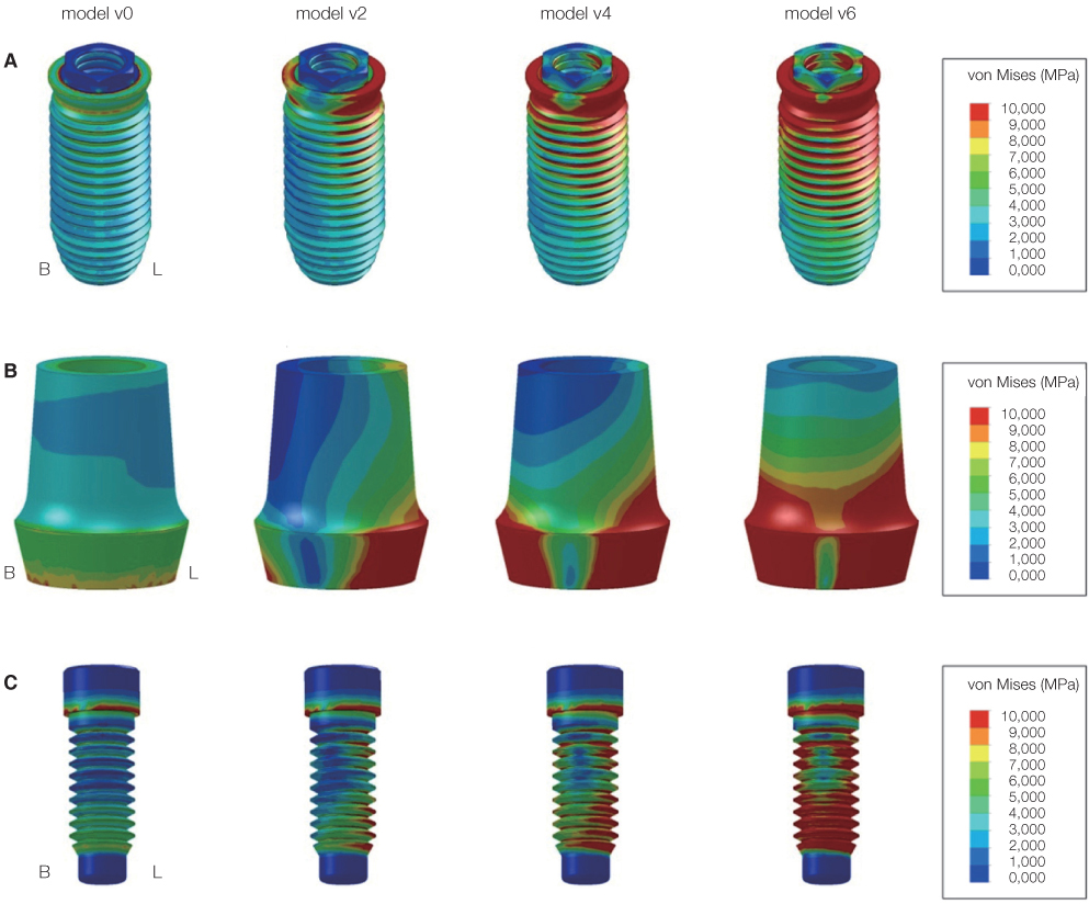
Fig. 6 Distribution of the von-Mises stress around implant components under vertical load. (A) Implant fixture, (B) abutment, (C) screw.

Fig. 7 Maximum von-Mises stress around cortical bone under oblique load.
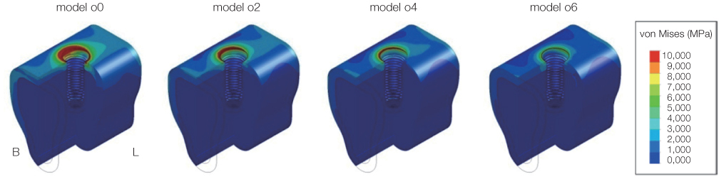
Fig. 8 Distribution of von-Mises stress around cortical bone under oblique load.

Fig. 9 Maximum von-Mises stress around implant components under oblique load. In case of model o4, o5, and o6, the stress decrement at the implant was larger than that at the abutment and screw. And the model with the lingual cantilever of 5 mm (model o5) was most affected.

Fig. 10 Distribution of the von-Mises stress around implant components under oblique load. (A) Implant fixture, (B) abutment, (C) screw.
Reference
-
1. Brånemark PI. Osseointegration and its experimental background. J Prosthet Dent. 1983; 50:399–410.2. Adell R, Lekholm U, Rockler B, Brånemark PI. A 15-year study of osseointegrated implants in the treatment of the edentulous jaw. Int J Oral Surg. 1981; 10:387–416.3. Brunski JB. Biomaterials and biomechanics in dental implant design. Int J Oral Maxillofac Implants. 1988; 3:85–97.4. Sones AD. Complications with osseointegrated implants. J Prosthet Dent. 1989; 62:581–585.5. Williams KR, Watson CJ, Murphy WM, Scott J, Gregory M, Sinobad D. Finite element analysis of fixed prostheses attached to osseointegrated implants. Quintessence Int. 1990; 21:563–570.6. van Steenberghe D. A retrospective multicenter evaluation of the survival rate of osseointegrated fixtures supporting fixed partial prostheses in the treatment of partial edentulism. J Prosthet Dent. 1989; 61:217–223.7. Quirynen M, Naert I, van Steenberghe D. Fixture design and overload influence marginal bone loss and fixture success in the Brånemark system. Clin Oral Implants Res. 1992; 3:104–111.8. Isidor F. Loss of osseointegration caused by occlusal load of oral implants. A clinical and radiographic study in monkeys. Clin Oral Implants Res. 1996; 7:143–152.9. Payant L, Williams JE, Zwemer JD. Survey of dental implant practice. J Oral Implantol. 1994; 20:50–58.10. Lekholm U, van Steenberghe D, Herrmann I, Bolender C, Folmer T, Gunne J, Henry P, Higuchi K, Laney WR, Lindén U. Osseointegrated Implants in the treatment of partially edentulous jaws: a prospective 5-year multicenter study. Int J Oral Maxillofac Implants. 1994; 9:627–635.11. Adell R, Eriksson B, Lekholm U, Brånemark PI, Jemt T. Long-term follow-up study of osseointegrated implants in the treatment of totally edentulous jaws. Int J Oral Maxillofac Implants. 1990; 5:347–359.12. Tallgren A. The continuing reduction of the residual alveolar ridges in complete denture wearers: a mixed-longitudinal study covering 25 years. J Prosthet Dent. 1972; 27:120–132.13. Atwood DA, Coy WA. Clinical, cephalometric, and densitometric study of reduction of residual ridges. J Prosthet Dent. 1971; 26:280–295.14. Hutton JE, Heath MR, Chai JY, Harnett J, Jemt T, Johns RB, McKenna S, McNamara DC, van Steenberghe D, Taylor R, et al. Factors related to success and failure rates at 3-year follow-up in a multicenter study of overdentures supported by Brånemark implants. Int J Oral Maxillofac Implants. 1995; 10:33–42.15. Stegaroiu R, Sato T, Kusakari H, Miyakawa O. Influence of restoration type on stress distribution in bone around implants: a three-dimensional finite element analysis. Int J Oral Maxillofac Implants. 1998; 13:82–90.16. Rangert B, Krogh PH, Langer B, Van Roekel N. Bending overload and implant fracture: a retrospective clinical analysis. Int J Oral Maxillofac Implants. 1995; 10:326–334.17. Weinstein AM, Klawitter JJ, Anand SC, Schuessler R. Stress analysis of porous rooted dental implants. J Dent Res. 1976; 55:772–777.18. Baik SH, Heo SJ. A three dimensional finite element analysis of stress distributions in implant-retained mandibular dentures with various retainers. Seoul National University;2008. Doctor's Thesis.19. Kenney R, Richards MW. Photoelastic stress patterns produced by implant-retained overdentures. J Prosthet Dent. 1998; 80:559–564.20. Benzing UR, Gall H, Weber H. Biomechanical aspects of two different implant-prosthetic concepts for edentulous maxillae. Int J Oral Maxillofac Implants. 1995; 10:188–198.21. Lum LB, Osier JF. Load transfer from endosteal implants to supporting bone: an analysis using statics. Part one: Horizontal loading. J Oral Implantol. 1992; 18:343–348.22. Sertgöz A, Güvener S. Finite element analysis of the effect of cantilever and implant length on stress distribution in an implant-supported fixed prosthesis. J Prosthet Dent. 1996; 76:165–169.23. Yokoyama S, Wakabayashi N, Shiota M, Ohyama T. The influence of implant location and length on stress distribution for three-unit implant-supported posterior cantilever fixed partial dentures. J Prosthet Dent. 2004; 91:234–240.24. Iplikçioğlu H, Akça K. Comparative evaluation of the effect of diameter, length and number of implants supporting three-unit fixed partial prostheses on stress distribution in the bone. J Dent. 2002; 30:41–46.25. Lum LB, Osier JF. Load transfer from endosteal implants to supporting bone: an analysis using statics. Part two: Axial loading. J Oral Implantol. 1992; 18:349–353.26. Weinberg LA. The biomechanics of force distribution in implant-supported prostheses. Int J Oral Maxillofac Implants. 1993; 8:19–31.27. Clelland NL, Lee JK, Bimbenet OC, Gilat A. Use of an axisymmetric finite element method to compare maxillary bone variables for a loaded implant. J Prosthodont. 1993; 2:183–189.28. Rangert B, Jemt T, Jörneus L. Forces and moments on Branemark implants. Int J Oral Maxillofac Implants. 1989; 4:241–247.29. Misch CE. Available bone and dental implant treatment plans. In : Micsh CE, editor. Contemporary implant dentistry. 2nd eds. St Louis; MO: Mosby;1999. p. 178–199.30. Richter EJ, Orschall B, Jovanovic SA. Dental implant abutment resembling the two-phase tooth mobility. J Biomech. 1990; 23:297–306.31. Borchers L, Reichart P. Three-dimensional stress distribution around a dental implant at different stages of interface development. J Dent Res. 1983; 62:155–159.32. Lavernia CJ, Cook SD, Weinstein AM, Klawitter JJ. An analysis of stresses in a dental implant system. J Biomech. 1981; 14:555–560.
- Full Text Links
-
- Actions
-
Cited
- CITED
-
- Close
- Share
-
- Similar articles
-
- Three-dimensional finite element analysis on stress distribution of the mandibular implant-supported cantilever prostheses depending on the designs
- Finite element analysis of the effect of cantilever and implant orientation on stress distribution in a mandibular implant-supported bar overdenture
- Finite Element Analysis of Stress Distribution on Telescopic System for Mandibular Implant Supported Overdenture
- Effect of number of implants and cantilever design on stress distribution in three-unit fixed partial dentures: A three-dimensional finite element analysis
- THREE DIMENSIONAL FINITE ELEMENT STRESS ANALYSIS OF THE MANDIBULAR CONDYLE DURING UNILATERAL CLENCHING
