Diabetes Metab J.
2015 Feb;39(1):46-50. 10.4093/dmj.2015.39.1.46.
Intravitreal Ranibizumab for Subfoveal Choroidal Neovascularization from Age-Related Macular Degeneration with Combined Severe Diabetic Retinopathy
- Affiliations
-
- 1Institute of Vision Research, Department of Ophthalmology, Yonsei University College of Medicine, Seoul, Korea.
- 2Department of Ophthalmology, Kangbuk Samsung Hospital, Sungkyunkwan University School of Medicine, Seoul, Korea. ssjeye@yahoo.co.kr
- 3Department of Ophthalmology, Korea University College of Medicine, Seoul, Korea.
- 4Department of Ophthalmology, Seoul National University College of Medicine, Seoul, Korea.
- KMID: 2280656
- DOI: http://doi.org/10.4093/dmj.2015.39.1.46
Abstract
- BACKGROUND
To evaluate the efficacy of intravitreal ranibizumab for subfoveal choroidal neovascularization (CNV) from age-related macular degeneration (AMD) with combined severe diabetic retinopathy (DR).
METHODS
This retrospective, interventional case series included eleven patients (mean age, 70.09 years; range, 54 to 83 years) with at least severe non-proliferative DR and subfoveal CNV secondary to AMD. Each subject was treated with intravitreal injections of 0.5 mg ranibizumab. The primary outcomes included change in best-corrected visual acuity and central subfield thickness (CST) on optical coherence tomography (OCT).
RESULTS
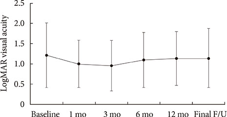
The mean follow-up time was 16.7+/-14 months (range, 6 to 31 months). Mean visual acuity improved from 1.21+/-0.80 logarithm of the minimum angle of resolution (logMAR) to 1.0+/-0.6 logMAR (P=0.107), 0.95+/-0.62 logMAR (P=0.044), 1.10+/-0.68 logMAR (P=0.296), and 1.13+/-0.66 logMAR (P=0.838) at 1, 3, 6, and 12 months after injection, respectively. Eight patients (72.7%) gained or maintained vision (mean 0.32 logMAR), whereas three patients (27.3%) lost more than one line of vision (mean 0.51 logMAR). The mean OCT CST was 343.9+/-134.6 microm at baseline, and the mean CST at 1, 3, 6, 12 months after the injection was 367.8+/-172.1 (P=0.864), 346.2+/-246.2 (P=0.857), 342+/-194.1 (P=0.551), and 294.2+/-108.3 microm (P=0.621), respectively.
CONCLUSION
Intravitreal ranibizumab injection can be considered to be a therapy for the stabilization of subfoveal CNV secondary to AMD with combined severe DR. However, these patients might exhibit limited visual improvement after treatment.
MeSH Terms
Figure
Reference
-
1. Pascolini D, Mariotti SP, Pokharel GP, Pararajasegaram R, Etya'ale D, Negrel AD, Resnikoff S. 2002 global update of available data on visual impairment: a compilation of population-based prevalence studies. Ophthalmic Epidemiol. 2004; 11:67–115.2. Congdon N, O'Colmain B, Klaver CC, Klein R, Munoz B, Friedman DS, Kempen J, Taylor HR, Mitchell P. Eye Diseases Prevalence Research Group. Causes and prevalence of visual impairment among adults in the United States. Arch Ophthalmol. 2004; 122:477–485.3. Yoon KC, Mun GH, Kim SD, Kim SH, Kim CY, Park KH, Park YJ, Baek SH, Song SJ, Shin JP, Yang SW, Yu SY, Lee JS, Lim KH, Park HJ, Pyo EY, Yang JE, Kim YT, Oh KW, Kang SW. Prevalence of eye diseases in South Korea: data from the Korea National Health and Nutrition Examination Survey 2008-2009. Korean J Ophthalmol. 2011; 25:421–433.4. Wild S, Roglic G, Green A, Sicree R, King H. Global prevalence of diabetes: estimates for the year 2000 and projections for 2030. Diabetes Care. 2004; 27:1047–1053.5. Antonetti DA, Klein R, Gardner TW. Diabetic retinopathy. N Engl J Med. 2012; 366:1227–1239.6. Mohan N, Monickaraj F, Balasubramanyam M, Rema M, Mohan V. Imbalanced levels of angiogenic and angiostatic factors in vitreous, plasma and postmortem retinal tissue of patients with proliferative diabetic retinopathy. J Diabetes Complications. 2012; 26:435–441.7. Frank RN, Amin RH, Eliott D, Puklin JE, Abrams GW. Basic fibroblast growth factor and vascular endothelial growth factor are present in epiretinal and choroidal neovascular membranes. Am J Ophthalmol. 1996; 122:393–403.8. Kvanta A, Algvere PV, Berglin L, Seregard S. Subfoveal fibrovascular membranes in age-related macular degeneration express vascular endothelial growth factor. Invest Ophthalmol Vis Sci. 1996; 37:1929–1934.9. Otani A, Takagi H, Oh H, Koyama S, Ogura Y, Matumura M, Honda Y. Vascular endothelial growth factor family and receptor expression in human choroidal neovascular membranes. Microvasc Res. 2002; 64:162–169.10. Brown DM, Kaiser PK, Michels M, Soubrane G, Heier JS, Kim RY, Sy JP, Schneider S. ANCHOR Study Group. Ranibizumab versus verteporfin for neovascular age-related macular degeneration. N Engl J Med. 2006; 355:1432–1444.11. Kaiser PK, Blodi BA, Shapiro H, Acharya NR. MARINA Study Group. Angiographic and optical coherence tomographic results of the MARINA study of ranibizumab in neovascular age-related macular degeneration. Ophthalmology. 2007; 114:1868–1875.12. Bhatnagar P, Spaide RF, Takahashi BS, Peragallo JH, Freund KB, Klancnik JM Jr, Cooney MJ, Slakter JS, Sorenson JA, Yannuzzi LA. Ranibizumab for treatment of choroidal neovascularization secondary to age-related macular degeneration. Retina. 2007; 27:846–850.13. Rosenfeld PJ, Brown DM, Heier JS, Boyer DS, Kaiser PK, Chung CY, Kim RY. MARINA Study Group. Ranibizumab for neovascular age-related macular degeneration. N Engl J Med. 2006; 355:1419–1431.14. Brown DM, Michels M, Kaiser PK, Heier JS, Sy JP, Ianchulev T. ANCHOR Study Group. Ranibizumab versus verteporfin photodynamic therapy for neovascular age-related macular degeneration: two-year results of the ANCHOR study. Ophthalmology. 2009; 116:57–65.15. Caldwell RB, Slapnick SM, McLaughlin BJ. Decreased anionic sites in Bruch’s membrane of spontaneous and drug-induced diabetes. Invest Ophthalmol Vis Sci. 1986; 27:1691–1697.16. Early Treatment Diabetic Retinopathy Study Research Group. Grading diabetic retinopathy from stereoscopic color fundus photograph: an extension of the modified Airlie House classification. ETDRS report number 10. Ophthalmology. 1991; 98:5 Suppl. 786–806.17. Barbazetto I, Burdan A, Bressler NM, Bressler SB, Haynes L, Kapetanios AD, Lukas J, Olsen K, Potter M, Reaves A, Rosenfeld P, Schachat AP, Strong HA, Wenkstern A. Treatment of Age-Related Macular Degeneration with Photodynamic Therapy Study Group. Verteporfin in Photodynamic Therapy Study Group. Photodynamic therapy of subfoveal choroidal neovascularization with verteporfin: fluorescein angiographic guidelines for evaluation and treatment: TAP and VIP report No. 2. Arch Ophthalmol. 2003; 121:1253–1268.18. Ladd BS, Solomon SD, Bressler NM, Bressler SB. Photodynamic therapy with verteporfin for choroidal neovascularization in patients with diabetic retinopathy. Am J Ophthalmol. 2001; 132:659–667.19. Diabetic Retinopathy Clinical Research Network. Elman MJ, Aiello LP, Beck RW, Bressler NM, Bressler SB, Edwards AR, Ferris FL 3rd, Friedman SM, Glassman AR, Miller KM, Scott IU, Stockdale CR, Sun JK. Randomized trial evaluating ranibizumab plus prompt or deferred laser or triamcinolone plus prompt laser for diabetic macular edema. Ophthalmology. 2010; 117:1064–1077.20. Yoshikawa T, Ogata N, Wada M, Otsuji T, Takahashi K. Characteristics of age-related macular degeneration in patients with diabetic retinopathy. Jpn J Ophthalmol. 2011; 55:235–240.
- Full Text Links
- Actions
-
Cited
- CITED
-
- Close
- Share
- Similar articles
-
- The Efficacy of Ranibizumab for Choroidal Neovascularization in Age-related Macular Degeneration
- Macular Hole Following Intravitreal Ranibizumab Injections for Choroidal Neovascularization
- Effect of Photodynamic Therapy and Intravitreal Triamcinolone Acetonide on Choroidal Neovascularization in Age-related Macular Degeneration
- Effects and Prognostic Factors of Intravitreal Bevacizumab Injection on Choroidal Neovascularization from Age-Related Macular Degeneration
- Macular Hole after Single Intravitreal Injection of Ranibizumab in a Patient with Age-Related Macular Degeneration


