Leiomyosarcoma of the Middle Ear and Temporal Bone
- Affiliations
-
- 1Department of Otorhinolaryngology, Yonsei University College of Medicine, Seoul, Korea. wsleemd@yuhs.ac
Abstract
- Leiomyosarcoma is a malignant tumor of smooth muscle cells that is exceedingly rare in the middle ear and temporal bone. Wide surgical resection is treatment of choice and adjuvant treatment has not proven to be of benefit. This is a report on a patient with otorrhea and rapidly growing mass on postauricualr area. A tumor that was mainly located in the middle ear and temporal bone was surgically removed and proved to be a leiomyosarcoma. The optimal surgical technique and other treatment strategy are discussed.
Keyword
Figure
-
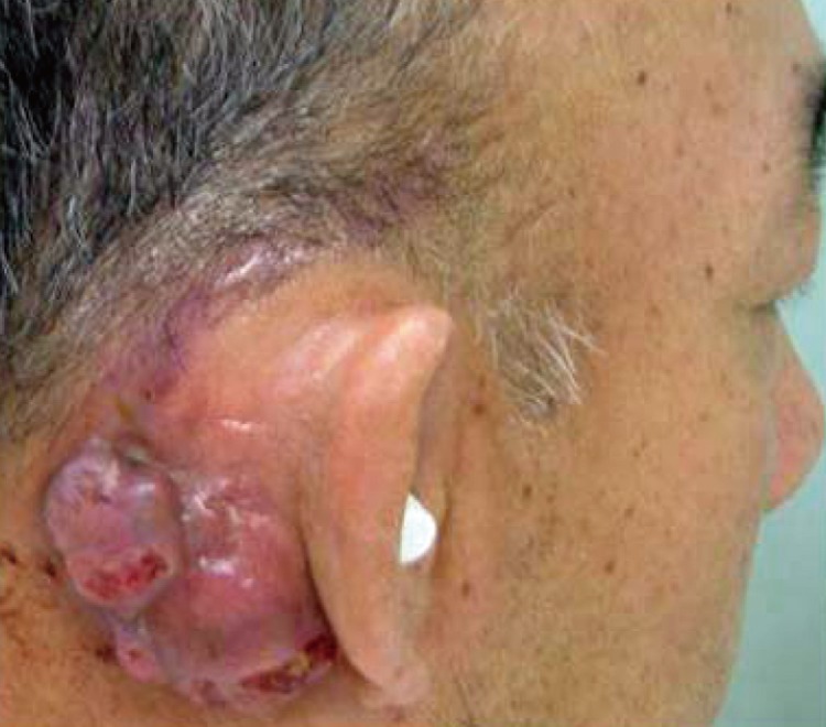
Fig. 1 The exophytic and polypoid mass about 5×4 cm in the right postauricualr region. There was a scar of the previous surgical incision on the overlying skin. The mass was reddish and the surrounding skin had color alteration.
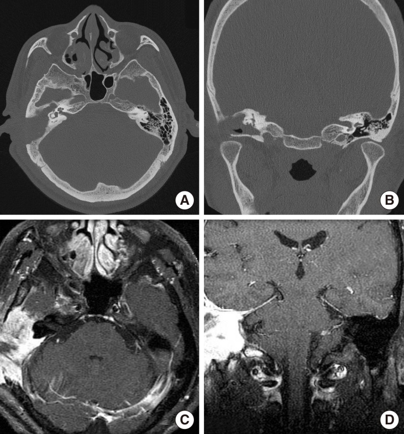
Fig. 2 Leiomyosarcoma of middle ear and temporal bone, (A) axial, (B) coronal computed tomography sections. A massive destructive lesion involves the temporal bone and the partial petrous bone with preservation of inner ear. (C) T1-weighted axial, (D) T1-weighted coronal magnetic resonance sections. The tumor spreads to pterygoid muscles, posterior margin of mandible and middle and posterior cranial fossa dura.
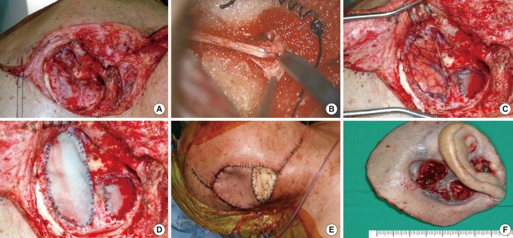
Fig. 3 The surgical procedure of leiomyosarcoma of the middle ear and temporal bone. (A) The excisions of the temporal bone was conducted including the external auditory canal, the middle ear, the vestibular organ, middle and posterior fossa dura, and the partial petrous bone, and the cochlear, the lateral sinus, and the jugular bulb remain intact. (B) The delicate epineural dissection of facial nerve was performed from tympanic segment to stylomastoid foramen. (C) The resections of the dura located on the middle and posterior cranial fossa were made. (D) The repair of the dura was done with Lyodura. (E) The approximation of the pectoralis muscle flap was done. (F) The specimen after en-bloc resection.
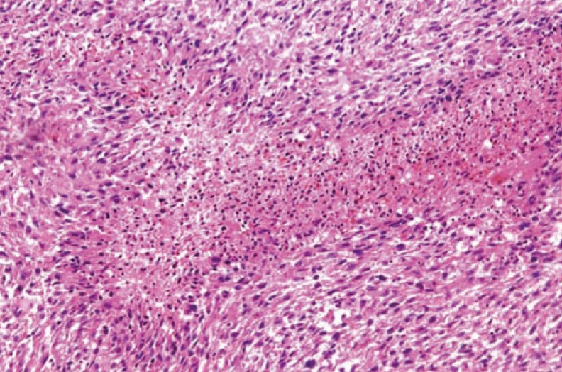
Fig. 4 The histology of leiomyosarcoma. It shows composed of spindle cells with cigar-shaped nuclei. There are mitotic activity and pleomorphic nuclei (H&E, ×100).

Fig. 5 The postoperative facial function. The facial nerve function was well preserved as House-Brackmann grade II after epineural dissection of facial nerve.
Reference
-
1. Zbaren P, Ruchti C. Leiomyosarcoma of the middle ear and temporal bone. Ann Otol Rhinol Laryngol. 1994; 7. 103(7):537–541. PMID: 8024216.
Article2. Mindell RS, Calcaterra TC, Ward PH. Leiomyosarcoma of the head and neck: a review of the literature and report of two cases. Laryngoscope. 1975; 5. 85(5):904–910. PMID: 1142965.
Article3. Marioni G, Bertino G, Mariuzzi L, Bergamin-Bracale AM, Lombardo M, Beltrami CA. Laryngeal leiomyosarcoma. J Laryngol Otol. 2000; 5. 114(5):398–401. PMID: 10912277.
Article4. Mankin HJ, Casas-Ganem J, Kim JI, Gebhardt MC, Hornicek FJ, Zeegen EN. Leiomyosarcoma of somatic soft tissues. Clin Orthop Relat Res. 2004; 4. (421):225–231. PMID: 15123952.
Article5. Farshid G, Pradhan M, Goldblum J, Weiss SW. Leiomyosarcoma of somatic soft tissues: a tumor of vascular origin with multivariate analysis of outcome in 42 cases. Am J Surg Pathol. 2002; 1. 26(1):14–24. PMID: 11756765.6. Eilber FR, Eckardt J. Surgical management of soft tissue sarcomas. Semin Oncol. 1997; 10. 24(5):526–533. PMID: 9344318.7. Yang JC, Chang AE, Baker AR, Sindelar WF, Danforth DN, Topalian SL, et al. Randomized prospective study of the benefit of adjuvant radiation therapy in the treatment of soft tissue sarcomas of the extremity. J Clin Oncol. 1998; 1. 16(1):197–203. PMID: 9440743.
Article8. Suit HD, Mankin HJ, Wood WC, Gebhardt MC, Harmon DC, Rosenberg A, et al. Treatment of the patient with stage M0 soft tissue sarcoma. J Clin Oncol. 1988; 5. 6(5):854–862. PMID: 3367188.
Article9. Cheng EY, Dusenbery KE, Winters MR, Thompson RC. Soft tissue sarcomas: preoperative versus postoperative radiotherapy. J Surg Oncol. 1996; 2. 61(2):90–99. PMID: 8606553.
Article10. Akcam T, Oysul K, Birkent H, Gerek M, Yetiser S. Leiomyosarcoma of the head and neck: report of two cases and review of the literature. Auris Nasus Larynx. 2005; 6. 32(2):209–212. PMID: 15917182.
Article11. Ogawa A, Sando I. Spatial occupancy of vessels and facial nerve in the facial canal. Ann Otol Rhinol Laryngol. 1982; Jan-Feb. 91(1 Pt 1):14–19. PMID: 7073171.
Article12. Gal TJ, Kerschner JE, Futran ND, Bartels LJ, Farrior JB, Ridley MB, et al. Reconstruction after temporal bone resection. Laryngoscope. 1998; 4. 108(4 Pt 1):476–481. PMID: 9546255.
Article
- Full Text Links
-
- Actions
-
Cited
- CITED
-
- Close
- Share
-
- Similar articles
-
- Osteoma of the Promontory Mimicking a Congenital Cholesteatoma
- Aberrant Internal Carotid Artery in the Middle Ear: A Case Report
- Clinical Features of Cholesterol Granuloma in Temporal Bone
- A Case of Middle Ear Lipoma
- Effects of middle ear and temporal bone pathology on bone conduction hearing level in chronic otitis media
