Bipolar Intra-articular Radiofrequency Thermocoagulation of the Thoracic Facet Joints: A Case Series of a New Technique
- Affiliations
-
- 1Department of Anesthesiology, Division of Pain Medicine, Henry Ford Medical Center, Detroit, USA. Dkim1@hfhs.org
- KMID: 2278201
- DOI: http://doi.org/10.3344/kjp.2014.27.1.43
Abstract
- BACKGROUND
This study tests the hypothesis that of bipolar radiofrequency thermocoagulation of the thoracic facet joint capsule may provide a safe and effect method of pain control from thoracic facet origin.
METHODS
Among patients suffering from localized mid back pain, nine patients with thoracic facet disease confirmed by magnetic resonance image and diagnostic thoracic facet block were enrolled. Bipolar radiofrequency ablation in the inferior aspect of the thoracic facet joint was done. Visual Analog Scale (VAS) was measured pre-intervention and 1 month post-intervention. Any complications and changes in amount of pain medication were recorded.
RESULTS
Significant 47.6% reduction in VAS was noted at 1 month. There were no serious complications.
CONCLUSIONS
Intra-articular bipolarradiofrequency thermocoagulation of the thoracic facet joint may be a technically easier and valid method of treating mid back pain of thoracic facet origin.
Keyword
MeSH Terms
Figure
-
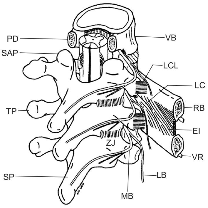
Fig. 1 Illustration of medial branch (MB) and lateral branch (LB) thoracic spine. Bogduk N (ed). Practice Guidelines for Spinal Diagnostics and Treatment Procedures. International Spine Injection Society (ISIS) 2004. Reproduced with permission of the publishers. PD: pedicle, VB: body of vertebra, SAP: superior articular process, LCL: lateral costovertebral ligament, LC: levator costorum, TP: transverse process, RB: rib, EI: external intercostal muscle, ZJ: zygaphophysial joint, VR: ventral root, SP: spinous process.
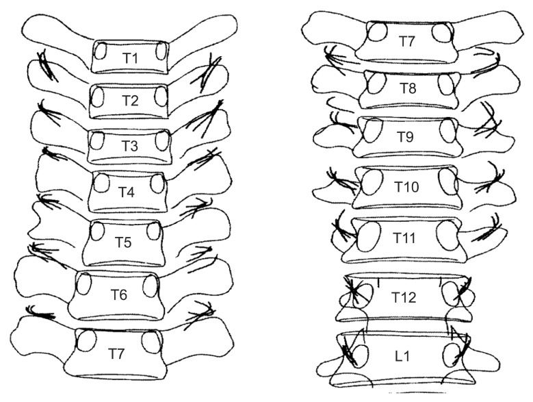
Fig. 2 Illustration of variation of position of medial branch in thoracic spine. Bogduk N (ed). Practice Guidelines for Spinal Diagnostics and Treatment Procedures,. International Spine Injection Society (ISIS) 2004. Reproduced with permission of the publishers.
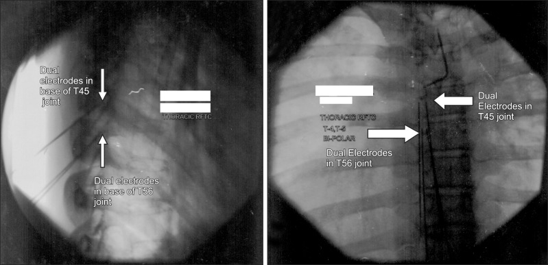
Fig. 3 Lateral and AP views of bipolar RFTC at left T4-5 and T5-6 facet joints. Note pairs of electrodes side by side approximately 5 mm apart in the inferior aspect of each joint.
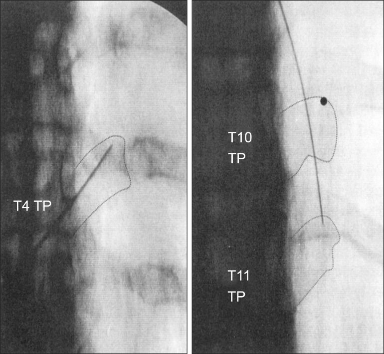
Fig. 4 Medial branch block T3 and T10 at the lateral end of superior border of the transverse process using opposite oblique orientation to see the superior tip of the transverse process: the "Pinnochio" view. Bogduk N (ed). Practice Guidelines for Spinal Diagnostics and Treatment Procedures. International Spine Injection Society (ISIS) 2004. Reproduced with permission of the publishers.
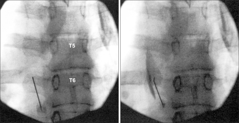
Fig. 5 Medial branch block left T5 above superior aspect of tip of left T6 transverse process. Bogduk N (ed). Practice Guidelines for Spinal Diagnostics and Treatment Procedures. International Spine Injection Society (ISIS) 2004. Reproduced with permission of the publishers.
Cited by 2 articles
-
Clinical Effectiveness of Ultrasound-guided Costotransverse Joint Injection in Thoracic Back Pain Patients
Kyung Bong Yoon, Shin Hyung Kim, Sang Jun Park, Ji Ae Moon, Duck Mi Yoon
Korean J Pain. 2016;29(3):197-201. doi: 10.3344/kjp.2016.29.3.197.Ultrasound-guided interventions for controlling the thoracic spine and chest wall pain: a narrative review
Donghwi Park, Min Cheol Chang
J Yeungnam Med Sci. 2022;39(3):190-199. doi: 10.12701/jyms.2022.00192.
Reference
-
1. Linton SJ, Hellsing AL, Halldén K. A population-based study of spinal pain among 35-45-year-old individuals. Prevalence, sick leave, and health care use. Spine (Phila Pa 1976). 1998; 23:1457–1463. PMID: 9670397.2. Wilson PR. Thoracic facet joint syndrome - a clinical entity? Pain. 1987; 30(Suppl 1):S87.
Article3. Stilwell DL Jr. The nerve supply of the vertebral column and its associated structures in the monkey. Anat Rec. 1956; 125:139–169. PMID: 13354975.
Article4. Chua WH, Bogduk N. The surgical anatomy of thoracic facet denervation. Acta Neurochir (Wien). 1995; 136:140–144. PMID: 8748844.
Article5. Bogduk N. International Spine Intervention Society, Standards Committee. Thoracic medial branch blocks. Practice guidelines for spinal diagnostic and treatment procedures. San Francisco (CA): International Spine Intervention Society;2004. p. 330–347.6. Tzaan WC, Tasker RR. Percutaeous radiofrequency facet rhizotomy--experience with 118 procdedures and reappraisal of its value. Can J Neurol Sci. 2000; 27:125–130. PMID: 10830345.
Article7. Stolker RJ, Vervest AC, Groen GJ. Percutaneous facet denervation in chronic thoracic spinal pain. Acta Neurochir (Wien). 1993; 122:82–90. PMID: 8333313.
Article8. Ferrante FM, King LF, Roche EA, Kim PS, Aranda M, Delaney LR, et al. Radiofrequency sacroiliac joint denervation for sacroiliac syndrome. Reg Anesth Pain Med. 2001; 26:137–142. PMID: 11251137.
Article9. Wedley JR, Gauci CA. Handbook of clinical techniques in the management of chronic pain. Sacroiliac joint denervation. Langhorne (PA): Harwood Academic;1994. p. 12–13.10. Manchikanti L, Singh V, Pampati V, Beyer CD, Damron KS. Evaluation of the prevalence of facet joint pain in chronic thoracic pain. Pain Physician. 2002; 5:354–359. PMID: 16886012.
Article11. Manchikanti L, Boswell MV, Singh V, Pampati V, Damron KS, Beyer CD. Prevalence of facet joint pain in chronic spinal pain of cervical, thoracic, and lumbar regions. BMC Musculoskelet Disord. 2004; 5:15. PMID: 15169547.
Article12. Manchikanti L, Singh V, Falco FJ, Cash KA, Pampati V. Effectiveness of thoracic medial branch blocks in managing chronic pain: a preliminary report of a randomized, double-blind controlled trial. Pain Physician. 2008; 11:491–504. PMID: 18690278.13. Manchikanti L, Manchikanti KN, Manchukonda R, Pampati V, Cash KA. Evaluation of therapeutic thoracic medial branch block effectiveness in chronic thoracic pain: a prospective outcome study with minimum 1-year follow up. Pain Physician. 2006; 9:97–105. PMID: 16703969.14. Stolker RJ, Vervest AC, Ramos LM, Groen GJ. Electrode positioning in thoracic percutaneous partial rhizotomy: an anatomical study. Pain. 1994; 57:241–251. PMID: 8090519.
Article15. Dreyfuss P, Tibiletti C, Dreyer S, Sobel J. Thoracic zygapophyseal joint pain: a review and description of an intraarticular block technique. Pain Digest. 1994; 4:46–54.16. Bogduk N, April C, Derby R. Diagnostic blocks of the synovial joints. In : White AH, editor. Spine care Vol. 1 Diagnosis and conservative treatment. St. Louis: Mosby;1995. p. 298–321.17. International Spine Intervention Society, Standards Committee. Bogduk N, editor. Practice guidelines for spinal diagnostic and treatment procedures. San Francisco (CA): International Spine Intervention Society;2004. p. 314–329.18. Dreyfuss P, Tibiletti C, Dreyer SJ. Thoracic zygapophyseal joint pain patterns. A study in normal volunteers. Spine (Phila Pa 1976). 1994; 19:807–811. PMID: 8202799.19. Fukui S, Ohseto K, Shiotani M. Patterns of pain induced by distending the thoracic zygapophyseal joints. Reg Anesth. 1997; 22:332–336. PMID: 9223198.
Article20. Fortin JD, McKee MJ. Thoracic facet blocks: bent needle technique. Pain Physician. 2003; 6:513–516. PMID: 16871307.
Article
- Full Text Links
-
- Actions
-
Cited
- CITED
-
- Close
- Share
-
- Similar articles
-
- Symptom Recurrence Pattern after Radiofrequency Thermocoagulationfor Facet Joint Syndrome
- Radiofrequency Facet Denervation for Low Back Pain: a Preliminary Report
- Radiofrequency Lesion Generation of the Articular Branches of the Obturator and Femoral Nerve for Hip Joint Pain: A case report
- Short Term Outcomes and Prognostic Factors Based on Radiofrequency Thermocoagulation on Lumbar Medial Branches
- Radiofrequency Facet Joint Denervation in the Treatment of Low Back Pain: Relationship with the Diagnostic Block
