A case of ovarian leiomyoma treated with laparoscopic mass excision
- Affiliations
-
- 1Department of Obstetrics and Gynecology, CHA Gangnam Medical Center, CHA University College of Medicine, Seoul, Korea. mila76@naver.com
- 2Department of Pathology, CHA Gangnam Medical Center, CHA University College of Medicine, Seoul, Korea.
- KMID: 2274132
- DOI: http://doi.org/10.5468/KJOG.2012.55.3.218
Abstract
- Primary ovarian leiomyoma is a rare tumor, accounting for only 0.5%-1% of benign ovarian neoplasms. About 80 cases have been reported in the literature worldwide to date. Most cases are asymptomatic and usually found incidentally in routine gynecologic workup, during surgery, or at autopsy. Ovarian leiomyoma is difficult to be differentiated from other ovarian benign and malignant neoplasms, particularly from large ovarian solid tumors. It is important to consider ovary-preserving surgery as the first treatment modality in patients of reproductive age after excluding the possibility of malignancy, given that 80% of ovarian leiomyomas occur in premenopausal women and the size of the mass is larger in younger patients. Here we report a case of ovarian leiomyoma in a 38-year-old female patient who underwent laparoscopic excision of left ovarian mass and uterine myomectomy.
Keyword
MeSH Terms
Figure
-
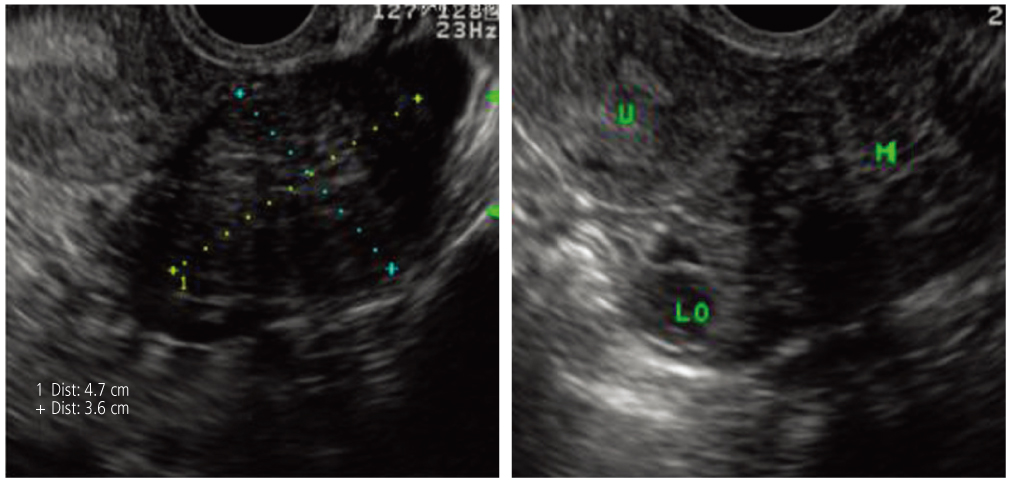
Fig. 1 Findings of transvaginal ultrasound. About 4.7 × 3.6 cm sized low mixed echo solid mass was noted in left ovary. U, uterus; LO, left ovary; M, mass.
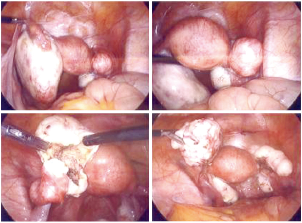
Fig. 2 Laparoscopic findings of left ovarian mass and subserosal myoma. After excision of ovarian mass, normal ovarian tissue was remained.
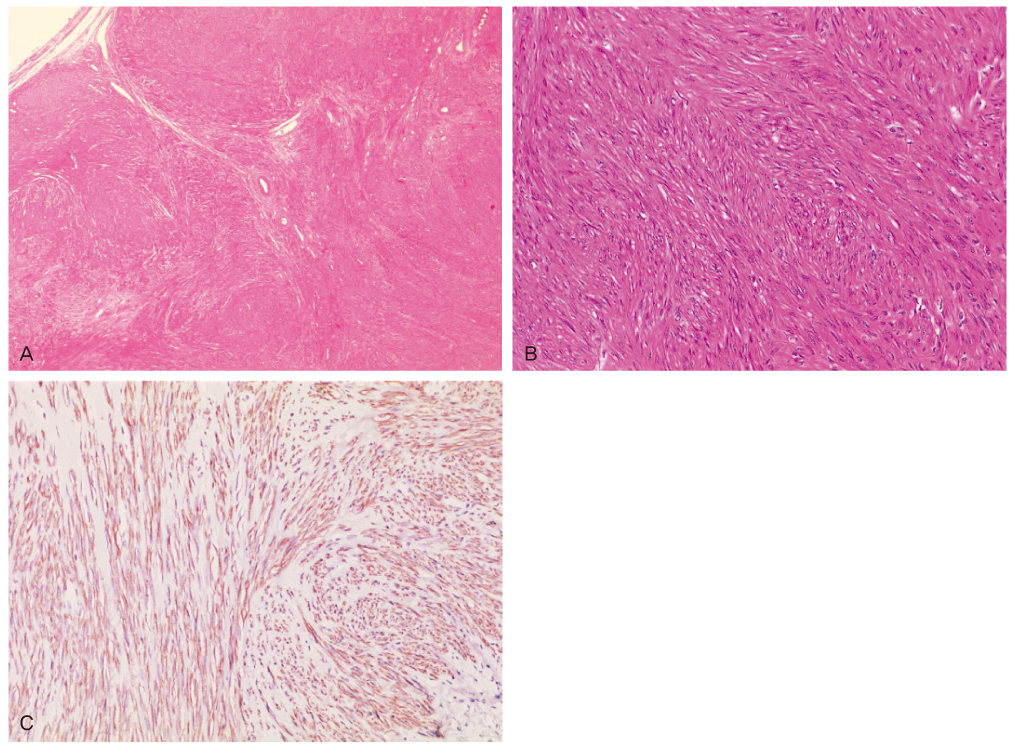
Fig. 3 Miscroscopic finding of the laparoscopically obtained ovarian tumor. (A) It was solid with whorling bundles of smooth muscle cells. The outer capsule of the mass is thin and the surrounding normal ovarian tissue was not included in the specimen (H&E, ×40). (B) Intertwining fascicles of benign smooth muscle cells with spindle-shaped, bland nuclei and slender eosinophilic cytoplasms are noted (H&E, ×200). (C) The tumor cells are strongly positive to desmin (desmin, ×200).
Reference
-
1. Doss BJ, Wanek SM, Jacques SM, Qureshi F, Ramirez NC, Lawrence WD. Ovarian leiomyomas: clinicopathologic features in fifteen cases. Int J Gynecol Pathol. 1999. 18:63–68.2. Wei C, Lilic N, Shorter N, Garrow E. Primary ovarian leiomyoma: a rare cause of ovarian tumor in adolescence. J Pediatr Adolesc Gynecol. 2008. 21:33–36.3. Koo YJ, Cho YJ, Kim JY, Lee JE, Kim ML, Kim JM, et al. Ovarian leiomyoma as a potential cause of compromised fertility. Fertil Steril. 2011. 95:1120.e11–1120.e14.4. Matamala MF, Nogales FF, Aneiros J, Herraiz MA, Caracuel MD. Leiomyomas of the ovary. Int J Gynecol Pathol. 1988. 7:190–196.5. Scully RE, Young RH, Clement PB. Atlas of tumor pathology: tumors of the ovary, maldeveloped gonads, fallopian tube, and broad ligament. 1998. Washington, DC: Armed Force Institute of Pathology.6. Khaffaf N, Khaffaf H, Wuketich S. Giant ovarian leiomyoma as a rare cause of acute abdomen and hydronephrosis. Obstet Gynecol. 1996. 87:872–873.7. Kurai M, Shiozawa T, Noguchi H, Konishi I. Leiomyoma of the ovary presenting with Meigs' syndrome. J Obstet Gynaecol Res. 2005. 31:257–262.8. San Marco L, Londero F, Stefanutti V, Costa L, Rocco M. Ovarian leiomyoma. Case report. Clin Exp Obstet Gynecol. 1991. 18:145–148.9. Fallahzadeh H, Dockerty MB, Lee RA. Leiomyoma of the ovary: report of five cases and review of the literature. Am J Obstet Gynecol. 1972. 113:394–398.10. Daniel Y, Lessing JB, Bar-Am A, Kupferminc MJ, Jossiphov J, Peyser MR. Treatment of bilateral multiple primary ovarian leiomyomas during pregnancy by way of conservative surgery: a case report. Eur J Obstet Gynecol Reprod Biol. 1997. 74:125–126.11. Tamada T, Sone T, Tanimoto D, Higashi H, Miyoshi H, Egashira N, et al. MRI appearance of primary giant ovarian leiomyoma in a hysterectomised woman. Br J Radiol. 2006. 79:e126–e128.12. Tomas D, Lenicek T, Tuckar N, Puljiz Z, Ledinsky M, Kruslin B. Primary ovarian leiomyoma associated with endometriotic cyst presenting with symptoms of acute appendicitis: a case report. Diagn Pathol. 2009. 4:25.13. Erkaya S, Kutlay B, Uygur D, Kara F, Tezer A. Primary ovarian leiomyoma in a postmenopausal woman. Acta Obstet Gynecol Scand. 2000. 79:79–87.14. Prayson RA, Hart WR. Primary smooth-muscle tumors of the ovary. A clinicopathologic study of four leiomyomas and two mitotically active leiomyomas. Arch Pathol Lab Med. 1992. 116:1068–1071.
