Korean J Obstet Gynecol.
2012 Mar;55(3):187-191. 10.5468/KJOG.2012.55.3.187.
A case of malignant fibrothecoma of the ovary
- Affiliations
-
- 1Department of Obstetrics and Gynecology, Dankook University College of Medicine, Cheonan, Korea. parkdkog@naver.com
- KMID: 2274125
- DOI: http://doi.org/10.5468/KJOG.2012.55.3.187
Abstract
- Fibrothecomas are mesenchymal tumors deriving from the ovarian stromal and consisting of theca-like elements and fibrous tissue. They are common, but their malignant counterpart is extraordinarily rare. Classical malignant fibrothecomas are said to show four or more mitotic figures per 10 high power fields. We have experienced a rare case of malignant ovarian fibrothecoma in a 72-year-old postmenopausal woman. We report a case with brief review of literature.
Keyword
Figure
-
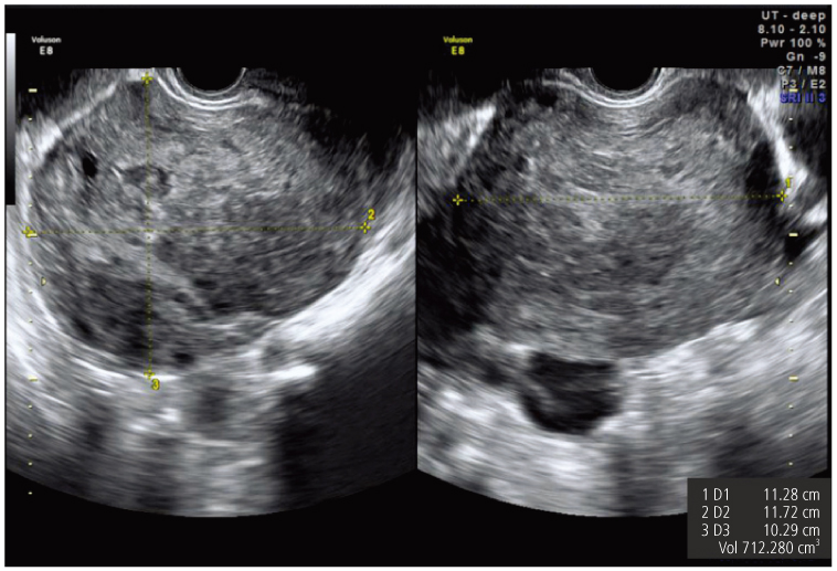
Fig. 1 Well defined heterogenous echoic solid mass (11.3×11.7×10.2 cm) with some tiny cystic changes abuting on atrophic uterus in right adnexa.
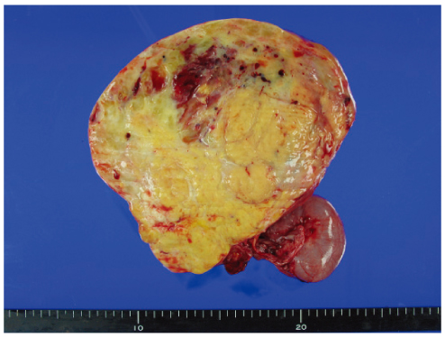
Fig. 2 The specimen received for frozen section consists of a circumscribed ovarian mass (16×16 cm), which is composed of yellow-gray solid firm mass with focal myxoid change.

Fig. 3 The tumor is composed of cellular, oval to spindle cells with pale cytoplasm and frequent mitosis (arrows) (H&E,×400).
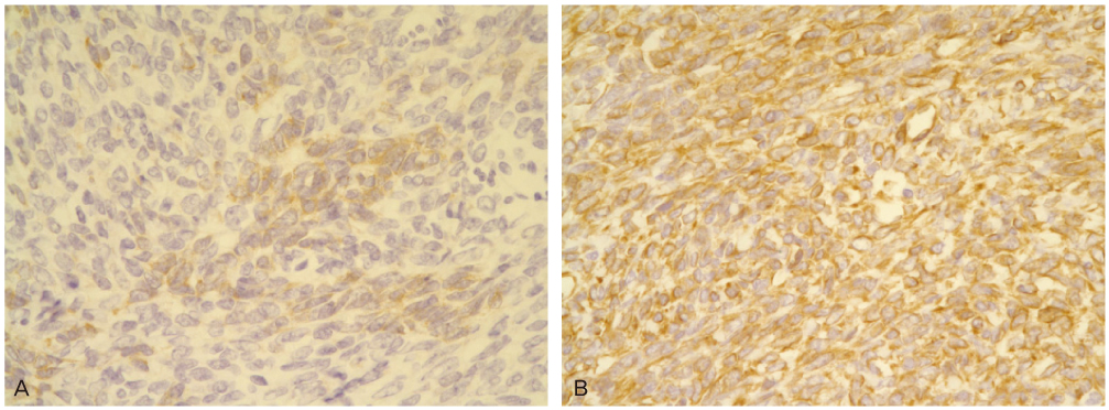
Fig. 4 The tumor cells are positive for vimentin (A) and alpha-inhibin (B). (Immunohistochemistry,×400).
Reference
-
1. Waxman M, Vuletin JC, Urcuyo R, Belling CG. Ovarian lowgrade stromal sarcoma with thecomatous features: a critical reappraisal of the so-called "malignant thecoma". Cancer. 1979. 44:2206–2217.2. Laufer L, Barki Y, Mordechai Y, Maor E, Mares A. Ovarian fibroma in a prepubertal girl. Pediatr Radiol. 1996. 26:40–42.3. Mawad NM, Hassanein OM. Ovarian fibro-thecoma in a 19 years old Sudanese girl. Gynaecological case report. Clin Exp Obstet Gynecol. 1994. 21:243–245.4. Stage AH, Grafton WD. Thecomas and granulosa-theca cell tumors of the ovary: an analysis of 51 tumors. Obstet Gynecol. 1977. 50:21–27.5. Sivanesaratnam V, Dutta R, Jayalakshmi P. Ovarian fibroma: clinical and histopathological characteristics. Int J Gynaecol Obstet. 1990. 33:243–247.6. Siekierska-Hellmann M, Sworczak K, Babińska A, Wojtylak S. Ovarian thecoma with androgenic manifestations in a postmenopausal woman. Gynecol Endocrinol. 2006. 22:405–408.7. Conte M, Guariglia L, Benedetti Panici P, Scambia G, Rabitti C, Capelli A, et al. Ovarian fibrothecoma: sonographic and histologic findings. Gynecol Obstet Invest. 1991. 32:51–54.8. Mak CW, Tzeng WS, Chen CY. Computed tomography appearance of ovarian fibrothecomas with and without torsion. Acta Radiol. 2009. 50:570–575.9. Prat J, Scully RE. Cellular fibromas and fibrosarcomas of the ovary: a comparative clinicopathologic analysis of seventeen cases. Cancer. 1981. 47:2663–2670.10. Le Bouëdec G, Glowaczower E, de Latour M, Fondrinier E, Kauffmann P, Dauplat J. Demons-Meigs' syndrome A case of thecoma and ovarian fibroma. J Gynecol Obstet Biol Reprod (Paris). 1992. 21:651–654.11. Bradley JA, Hamilton DN, McWhinnie DL, Briggs JD, Junor BJ. Sclerosing peritonitis after CAPD. Lancet. 1983. 2:572–573.12. Brown P, Baddeley H, Read AE, Davies JD, McGarry J. Sclerosing peritonitis, an unusual reaction to a beta-adrenergic-blocking drug (practolol). Lancet. 1974. 2:1477–1481.13. Clement PB, Young RH, Hanna W, Scully RE. Sclerosing peritonitis associated with luteinized thecomas of the ovary. A clinicopathological analysis of six cases. Am J Surg Pathol. 1994. 18:1–13.14. Chiappa A, Zbar AP, Bertani E, Biffi R, Luca F, Crotti C, et al. Primary and recurrent retroperitoneal soft tissue sarcoma: prognostic factors affecting survival. J Surg Oncol. 2006. 93:456–463.15. Avancès C, Mottet N, Mahatmat A, Chapuis E, Serre I, Culine S. Prognostic factors for first recurrence in patients with retroperitoneal sarcoma. Urol Oncol. 2006. 24:94–96.
- Full Text Links
-
- Actions
-
Cited
- CITED
-
- Close
- Share
-
- Similar articles
-
- A case of ovarian fibrothecoma with Mature teratoma of the Ipsilateral Ovary and ovarian fibroma of the contralateral Ovary
- Torsion of Collision Tumor: Dermoid Cyst and Fibrothecoma with Postmenopausal Bleeding
- A case of Meigs' syndrome and sclerosing peritonitis associated with fibrothecoma of bilateral ovary
- Multiple Synchronous Lesions in the Genital Tract of a Female: A Rare Combination with Unrelated Histogenesis
- A Case of Meigs` Syndrome with Elevated Serum CA 125 Level
