Korean J Obstet Gynecol.
2011 Nov;54(11):707-711. 10.5468/KJOG.2011.54.11.707.
A case of embryonal rhabdomyosarcoma of the uterine cervix in a middle-aged woman
- Affiliations
-
- 1Division of Gynecologic Oncology, Department of Obstetrics and Gynecology, Yonsei University College of Medicine, Seoul, Korea. san1@yuhs.ac
- 2Department of Pathology, Yonsei University College of Medicine, Seoul, Korea.
- KMID: 2274111
- DOI: http://doi.org/10.5468/KJOG.2011.54.11.707
Abstract
- Embryonal rhabdomyosarcoma (RMS) of the cervix is rare and most commonly occurs in the late teens and early 20s. We report a case of cervical embryonal RMS in a 52-year-old woman. This patient presented with an abnormal vaginal bleeding for 2 months and a mass protruding from the introitus, measuring 7 x 6 cm. She underwent radical abdominal hysterectomy with bilateral pelvic lymph node dissection and radical vaginectomy. The final pathologic result was cervical RMS, consistent with the Intergroup RMS study group IIC. Immunohistochemistry was positive for desmin, myogenin, and myogenic diffentiation 1. The patient received conservative management in a convalescent hospital without adjuvant treatment due to cerebral hemorrhage and relapsed septic condition after surgery.
Keyword
MeSH Terms
Figure
-
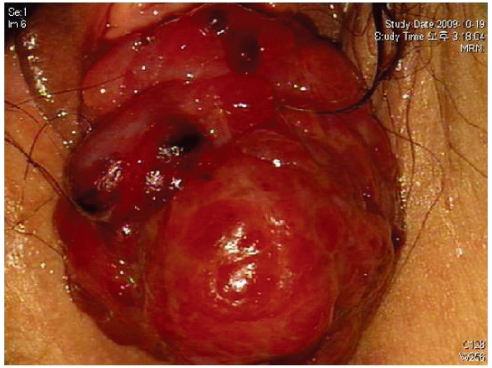
Fig. 1 A 7 × 6 cm size soft pinkish irregular mass arising from the uterine cervix and reaching up to the introitus.

Fig. 2 F-18 flurodeoxyglucose (FDG) positron emission tomography/ computed tomography finding. There was intense uptake in the protruding vaginal mass, consistent with malignancy. FDG uptake extension to the cervix area was seen, suggestive of malignant involvement and periregional infiltration was suspected.
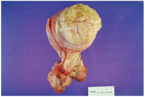
Fig. 3 Gross finding after surgery.
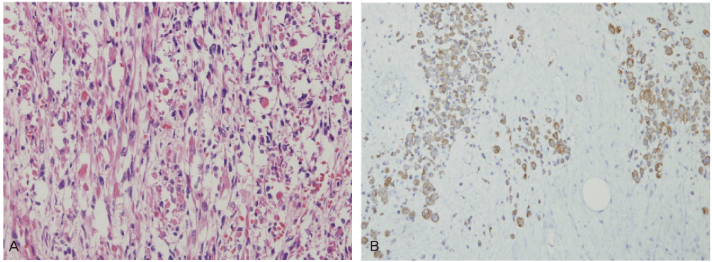
Fig. 4 (A) The tumor consists of oval to spindle shaped malignant embryonal rhabdomyoblasts with eosinophilic cytoplasm (H&E, ×200). (B) The desmin immunoreactivity confirms the myoblastic differentiation (×100).
Reference
-
1. Ghaemmaghami F, Karimi Zarchi M, Ghasemi M. Lower genital tract rhabdomyosarcoma: case series and literature review. Arch Gynecol Obstet. 2008. 278:65–69.2. Park JY, Jeo JS, Kim DY, Lee DH, Kim JH, Kim YM, et al. Two cases of embryonal rhabdomyosarcoma of the uterine cervix. Korean J Gynecol Oncol Colposc. 2002. 13:264–270.3. Newton WA Jr, Gehan EA, Webber BL, Marsden HB, van Unnik AJ, Hamoudi AB, et al. Classification of rhabdomyosarcomas and related sarcomas. Pathologic aspects and proposal for a new classification: an Intergroup Rhabdomyosarcoma Study. Cancer. 1995. 76:1073–1085.4. Hollowood K, Fletcher CD. Rhabdomyosarcoma in adults. Semin Diagn Pathol. 1994. 11:47–57.5. Ghavimi F, Mandell LR, Heller G, Hajdu SI, Exelby P. Prognosis in childhood rhabdomyosarcoma of the extremity. Cancer. 1989. 64:2233–2237.6. Zeisler H, Mayerhofer K, Joura EA, Bancher-Todesca D, Kainz C, Breitenecker G, et al. Embryonal rhabdomyosarcoma of the uterine cervix: case report and review of the literature. Gynecol Oncol. 1998. 69:78–83.7. Qualman S, Lynch J, Bridge J, Parham D, Teot L, Meyer W, et al. Prevalence and clinical impact of anaplasia in childhood rhabdomyosarcoma: a report from the Soft Tissue Sarcoma Committee of the Children's Oncology Group. Cancer. 2008. 113:3242–3247.8. Baiocchi G, Faloppa CC, Osório CA, Kumagai LY, Fukazawa EM, Cunha IW. Embryonal rhabdomyosarcoma of the uterine cervix in a 47-year-old woman. J Obstet Gynaecol Res. 2011. 37:940–946.9. Crist WM, Anderson JR, Meza JL, Fryer C, Raney RB, Ruymann FB, et al. Intergroup rhabdomyosarcoma study-IV: results for patients with nonmetastatic disease. J Clin Oncol. 2001. 19:3091–3102.10. Jang JB, Kang SH, Kim YT, Kim JW, No TW, Kim HG. A case of embryonal rhabdomyosarcoma of the uterine cervix in a 31 year old woman. Korean J Obstet Gynecol. 2001. 44:202–207.
- Full Text Links
-
- Actions
-
Cited
- CITED
-
- Close
- Share
-
- Similar articles
-
- A case of alveolar rhabdomyosarcoma of the uterine cervix
- A Case of Embryonal Rhabdomyosarcoma of the Uterine Cervix in a 31 Year Old Woman
- Two Cases of Embryonal Rhabdomyosarcoma of the Uterine Cervix
- Sarcoma botryoides (embryonal rhabdomyosarcoma) of the uterine cervix in sisters
- Embryonal Rhabdomyosarcoma of the Prostate
