A case of ovarian deciduosis in pregnancy
- Affiliations
-
- 1Department of Obstetrics and Gynecology, Yonsei University College of Medicine, Seoul, Korea. ywparkob@yuhs.ac
- KMID: 2274052
- DOI: http://doi.org/10.5468/KJOG.2011.54.7.373
Abstract
- Ectopic decidua (deciduosis) has been discovered in variable organs during pregnancy. Ovarian deciduosis, however, is a less frequent event during pregnancy. Ectopic decidua is a physiological phenomenon of pregnancy and arises from a progesterone-induced metaplasia of subserosal stromal cells. As we experienced a case of 21 weeks gestation who was diagnosed with ovarian deciduosis that was suspicious for ovarian malignant tumor, we present it with a brief review of literature.
Keyword
Figure
-

Fig. 1 (A) Transvaginal utrasonogram image shows a 10.9 × 3.9 cm sized heterogenous cystic mass with 6.6 × 3.5 cm, 2.5 × 1.4 cm and 1.8 × 1.4 cm of solid portion on left adnexa. (B) Doppler ultrasonogram image of the mass shows vascularization with pulsatility, a finding that is suggestive of a malignant mass (resistance index [RI], RI=0.37). PSV, peak systolic velocity; EDV, end-diastolic velocity; S/D, systolic:diastolic ratio.
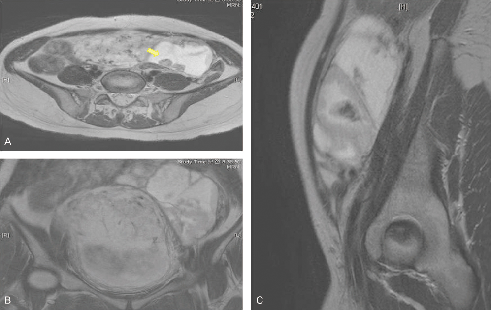
Fig. 2 Axial T2-weighted (A), coronal 2-weighted (B) and sagittal T2-weighted (C) images demonstrate late 2nd trimester pregnant state and about 10 × 7 × 4.8 cm sized mass at left adnexa. The solid component along the cyst wall shows intermediate to high signal intensity on T2-weighted images (T2WI). The nodule (arrow) with low signal intensity on T2WI is possibility of paramagnetic component such as hemosiderin. Twenty-one gestational week sized fetus is noted in (C).

Fig. 3 Histologic findings: ovary revealed a pinkish tan colored edematous stroma (A, H&E; ×40), deciduosis with hemosiderin laden macrophage (B, H&E; ×100).
Reference
-
1. Piccinni DJ, Spitale LS, Cabalier LR, Dionisio de Cabalier ME. Decidua in the peritoneal surface mimicking metastatic nodules. Findings during cesarean section. Rev Fac Cien Med Univ Nac Cordoba. 2002. 59:113–116.2. Fenjvesi A, Zivković S. Deciduosis peritonei: a case report. Med Pregl. 2005. 58:196–199.3. Szopiński TR, Sudoł-Szopińska I, Dzik T, Borówka A. Ectopic decidual reaction in the urinary bladder presenting as a vesical tumor. Urology. 2009. 74:1232–1233.4. Ghossain MA, Buy JN, Lignères C, Bazot M, Hassen K, Malbec L, et al. Epithelial tumors of the ovary: comparison of MR and CT findings. Radiology. 1991. 181:863–870.5. Troiano RN, McCarthy S. Magnetic resonance imaging evaluation of adnexal masses. Semin Ultrasound CT MR. 1994. 15:38–48.6. Büttner A, Bässler R, Theele C. Pregnancy-associated ectopic decidua (deciduosis) of the greater omentum. An analysis of 60 biopsies with cases of fibrosing deciduosis and leiomyomatosis peritonealis disseminata. Pathol Res Pract. 1993. 189:352–359.7. Tang LC, Cheung MY, Ma HK. Intraperitoneal bleeding from ectopic decidua following hormonal contraception. Case report. Br J Obstet Gynaecol. 1985. 92:102–103.8. Weller CV. The Ectopic Decidual Reaction and its Significance in Endometriosis. Am J Pathol. 1935. 11:287.1–290.1.9. Papp Z, Petri I, Villányi E, Tiszlavicz L, Ugocsai G. Deciduosis causing perforating appendicitis in the early postpartum period following caesarean section. Orv Hetil. 2008. 149:329–331.10. Shukla S, Pujani M, Singh SK. Ectopic decidual reaction mimicking peritoneal tubercles: a report of three cases. Indian J Pathol Microbiol. 2008. 51:519–520.
