Three-dimensional symmetry and parallelism of the skeletal and soft-tissue poria in patients with facial asymmetry
- Affiliations
-
- 1Department of Orthodontics, School of Dentistry, Dankook University, Cheonan, Korea. saegum@naver.com
- KMID: 2273428
- DOI: http://doi.org/10.4041/kjod.2014.44.2.62
Abstract
OBJECTIVE
The purpose of this study was to examine the symmetry and parallelism of the skeletal and soft-tissue poria by three-dimensional (3D) computed tomographic (CT) imaging.
METHODS
The locations of the bilateral skeletal and soft-tissue poria in 29 patients with facial asymmetry (asymmetric group) and 29 patients without facial asymmetry (symmetric group) were measured in 3D reconstructed models of CT images by using a 3D coordinate system. The mean intergroup differences in the anteroposterior and vertical angular deviations of the poria and their anteroposterior and vertical parallelism were statistically analyzed.
RESULTS
The symmetric and asymmetric groups showed significant anteroposterior angular differences in both the skeletal and the soft-tissue poria (p = 0.007 and 0.037, respectively; Mann-Whitney U-test). No significant differences in the anteroposterior and vertical parallelism of the poria were noted (p < or = 0.05; Wilcoxon signed-rank test).
CONCLUSIONS
In general, the skeletal poria are parallel to the soft-tissue poria. However, patients with facial asymmetry tend to have asymmetric poria.
Keyword
MeSH Terms
Figure
-
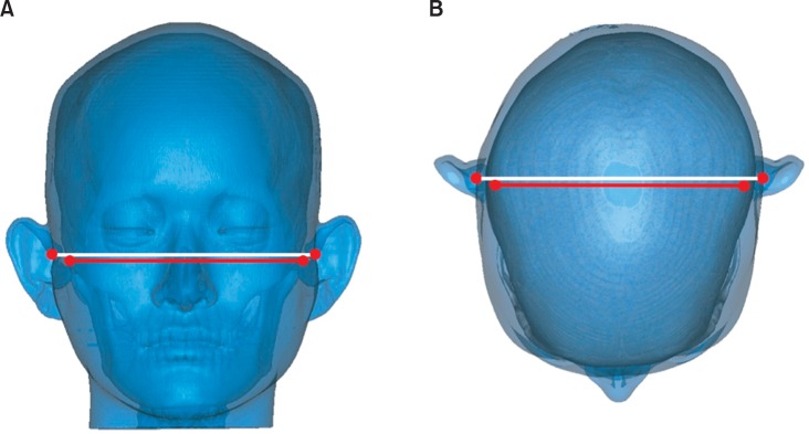
Figure 1 Parallelism of the skeletal and soft-tissue poria. A, Vertical parallelism; B, anteroposterior parallelism.
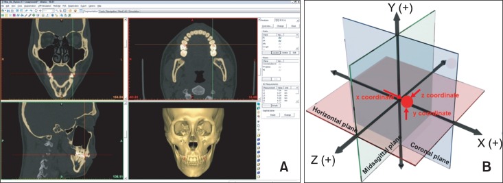
Figure 2 The three-dimensional (3D) reconstructed model. A, 3D reconstruction of computed tomographic images; B, 3D coordinate system.
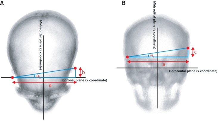
Figure 3 Measurement of the angular deviations. A, Anteroposterior deviation (θ1); B, vertical deviation (θ2).a, l x coordinate of left porion - x coordinate of right porion l; b, l z coordinate of left porion - z coordinate of right porion l; c, l y coordinate of left porion - y coordinate of right porion l.
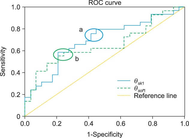
Figure 4 Receiver operating characteristic (ROC) curves of θsk1 and θsof1. a, cutoff section of θsk1; b, cutoff section of θsof1.
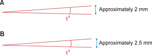
Figure 5 Conversion of angular deviation into linear deviation. A, For the skeletal poria; B, for the soft-tissue poria.
Cited by 1 articles
-
Cone-beam computed tomography based evaluation of rotational patterns of dentofacial structures in skeletal Class III deformity with mandibular asymmetry
Hyeong-Seok Ryu, Ki-Yong An, Kyung-Hwa Kang
Korean J Orthod. 2015;45(4):153-163. doi: 10.4041/kjod.2015.45.4.153.
Reference
-
1. Kim YH, Sato K, Mitani H, Shimizu Y, Kikuchi M. Asymmetry of the sphenoid bone and its suitability as a reference for analyzing craniofacial asymmetry. Am J Orthod Dentofacial Orthop. 2003; 124:656–662. PMID: 14666078.
Article2. Katsumata A, Fujishita M, Maeda M, Ariji Y, Ariji E, Langlais RP. 3D-CT evaluation of facial asymmetry. Oral Surg Oral Med Oral Pathol Oral Radiol Endod. 2005; 99:212–220. PMID: 15660095.
Article3. Park SH, Yu HS, Kim KD, Lee KJ, Baik HS. A proposal for a new analysis of craniofacial morphology by 3-dimensional computed tomography. Am J Orthod Dentofacial Orthop. 2006; 129:600.e23–600.e34. PMID: 16679198.
Article4. Kim WS, Lee KH, Hwang HS. Comparison of asymmetric degree between maxillofacial hard and soft tissue in facial asymmetric subjects using three-dimensional computed tomography. Korean J Orthod. 2005; 35:163–173.5. Ghom AG. Textbook of oral radiology. Noida, India: Elsevier (A Division of Reed Elsevier India Pvt. Limited);2008.6. Ahn JS, Hwang HS. Relationship between perception of facial asymmetry and posteroanterior cephalometric measurements. Korean J Orthod. 2001; 31:489–498.7. Lee MS, Chung DH, Lee JW, Cha KS. Assessing soft-tissue characteristics of facial asymmetry with photographs. Am J Orthod Dentofacial Orthop. 2010; 138:23–31. PMID: 20620830.
Article8. Kim KS, Kim YJ, Lee KH, Kim YH, Kook YA. Level of perception of changed lip protrusion and asymmetry of the lower facial height. Korean J Orthod. 2006; 36:434–441.9. Kwon TG, Park HS, Ryoo HM, Lee SH. A comparison of craniofacial morphology in patients with and without facial asymmetry--a three-dimensional analysis with computed tomography. Int J Oral Maxillofac Surg. 2006; 35:43–48. PMID: 15925488.
Article10. Yoo SK, Kim YO, Kim HJ, Kim NH, Jang YB, Kim KD, et al. Alignment of CT images of skull dysmorphology using anatomy-based perpendicular axes. Phys Med Biol. 2003; 48:2681–2695. PMID: 12974582.
Article11. Kitaura H, Yonetsu K, Kitamori H, Kobayashi K, Nakamura T. Standardization of 3-D CT measurements for length and angles by matrix transformation in the 3-D coordinate system. Cleft Palate Craniofac J. 2000; 37:349–356. PMID: 10912713.
Article12. Cho JH, Moon JY. Comparison of midsagittal reference plane in PA cephalogram and 3D CT. Korean J Orthod. 2010; 40:6–15.
Article13. Grayson BH, McCarthy JG, Bookstein F. Analysis of craniofacial asymmetry by multiplane cephalometry. Am J Orthod. 1983; 84:217–224. PMID: 6577794.
Article14. Peck S, Peck L, Kataja M. Skeletal asymmetry in esthetically pleasing faces. Angle Orthod. 1991; 61:43–48. PMID: 2012321.15. Maeda M, Katsumata A, Ariji Y, Muramatsu A, Yoshida K, Goto S, et al. 3D-CT evaluation of facial asymmetry in patients with maxillofacial deformities. Oral Surg Oral Med Oral Pathol Oral Radiol Endod. 2006; 102:382–390. PMID: 16920547.
Article16. Captier G, Leboucq N, Bigorre M, Canovas F, Bonnel F, Bonnafé A, et al. Plagiocephaly: morphometry of skull base asymmetry. Surg Radiol Anat. 2003; 25:226–233. PMID: 14504821.
Article17. Huang MH, Gruss JS, Clarren SK, Mouradian WE, Cunningham ML, Roberts TS, et al. The differential diagnosis of posterior plagiocephaly: true lambdoid synostosis versus positional molding. Plast Reconstr Surg. 1996; 98:765–774. discussion 775-6. PMID: 8823012.
Article18. Lo LJ, Marsh JL, Pilgram TK, Vannier MW. Plagiocephaly: differential diagnosis based on endocranial morphology. Plast Reconstr Surg. 1996; 97:282–291. PMID: 8559810.
Article19. Mulliken JB, Vander Woude DL, Hansen M, LaBrie RA, Scott RM. Analysis of posterior plagiocephaly: deformational versus synostotic. Plast Reconstr Surg. 1999; 103:371–380. PMID: 9950521.
Article20. Kane AA, Lo LJ, Vannier MW, Marsh JL. Mandibular dysmorphology in unicoronal synostosis and plagiocephaly without synostosis. Cleft Palate Craniofac J. 1996; 33:418–423. PMID: 8891373.
Article21. St John D, Mulliken JB, Kaban LB, Padwa BL. Anthropometric analysis of mandibular asymmetry in infants with deformational posterior plagiocephaly. J Oral Maxillofac Surg. 2002; 60:873–877. PMID: 12149730.
- Full Text Links
-
- Actions
-
Cited
- CITED
-
- Close
- Share
-
- Similar articles
-
- Analysis of Facial Asymmetry
- Facial asymmetry: a case report of localized linear scleroderma patient with muscular strain and spasm
- Comparison of asymmetric degree between maxillofacial hard and soft tissue in facial asymmetric subjects using three-dimensional computed tomography
- Evaluation of the mandibular asymmetry using the facial photographs and the radiographs
- Three-dimensional Assessment of Facial Soft Tissue after Orthognathic Surgery in Patients with Skeletal Class III and Asymmetry
