Three-dimensional assessment of upper lip positional changes according to simulated maxillary anterior tooth movements by white light scanning
- Affiliations
-
- 1Department of Orthodontics, School of Dentistry, Dankook University, Cheonan, Korea. selemos@naver.com
- KMID: 2273228
- DOI: http://doi.org/10.4041/kjod.2014.44.6.281
Abstract
OBJECTIVE
Esthetic improvements during orthodontic treatment are achieved by changes in positions of the lips and surrounding soft tissues. Facial soft-tissue movement has already been two-dimensionally evaluated by cephalometry. In this study, we aimed to three-dimensionally assess positional changes of the adult upper lip according to simulated maxillary anterior tooth movements by white light scanning.
METHODS
We measured changes in three-dimensional coordinates of labial landmarks in relation to maxillary incisor movements of normal adults simulated with films of varying thickness by using a white light scanner.
RESULTS
With increasing protraction, the upper lip moved forward and significantly upward. Labial movement was limited by the surrounding soft tissues. The extent of movement above the vermilion border was slightly less than half that of the teeth, showing strong correlation. Most changes were concentrated in the depression above the upper vermilion border. Labial movement toward the nose was reduced significantly.
CONCLUSIONS
After adequately controlling several variables and using white light scanning with high reproducibility and accuracy, the coefficient of determination showed moderate values (0.40-0.77) and significant changes could be determined. This method would be useful to predict soft-tissue positional changes according to tooth movements.
Figure
-

Figure 1 A, Normal occlusal position; B, film attachment.
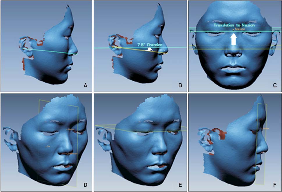
Figure 2 A, Camper's plane; B, true horizontal plane; C, translated horizontal plane; D, sagittal reference plane; E, axial reference plane; F, coronal reference plane.
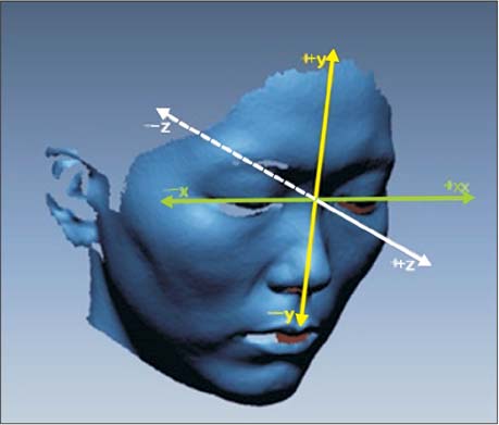
Figure 3 Three-dimensional coordinate system.
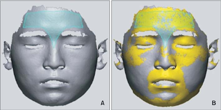
Figure 4 A, Forehead mesh region; B, best-fit alignment with forehead mesh region.
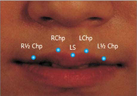
Figure 5 Labial landmarks. See Table 1 for their definitions.

Figure 6 Curves and points. See Table 1 for their definitions.
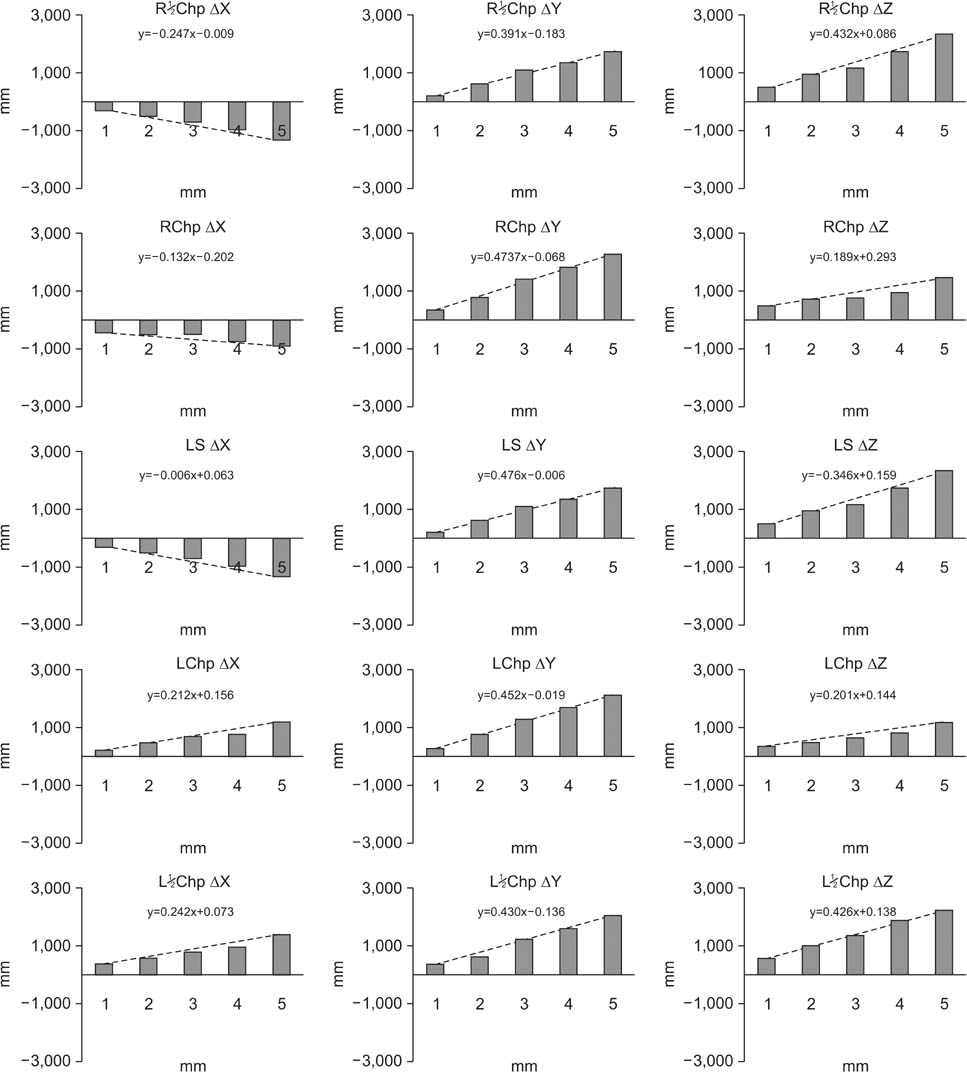
Figure 7 Results of simple linear regression of positional changes in labial landmarks according to film thickness. See Table 1 and Figure 5 for the definitions of landmarks.

Figure 8 Three-dimensional morphologic changes in Curve 1. The curves were divided into 11 sections, and ten equidistant points were added on each curve. RIPP, Right inferior philtrum point; MPP, most protrusive point.

Figure 9 Three-dimensional morphologic changes in Curve 2. The curves were divided into 11 sections, and ten equidistant points were added on each curve. Sn, Subnasale; MPP, most protrusive point.
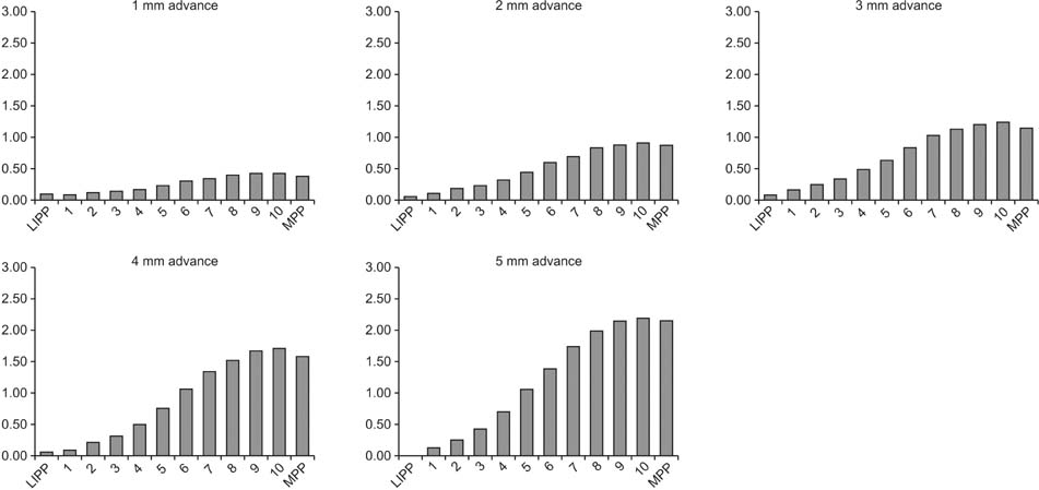
Figure 10 Three-dimensional morphologic changes in Curve 3. The curves were divided into 11 sections, and ten equidistant points were added on each curve. LIPP, Left inferior philtrum point; MPP, most protrusive point.
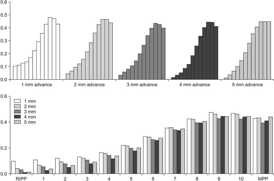
Figure 11 Soft-tissue movement ratio of Curve 1. The curves were divided into 11 sections, and ten equidistant points were added on each curve. RIPP, Right inferior philtrum point; MPP, most protrusive point.
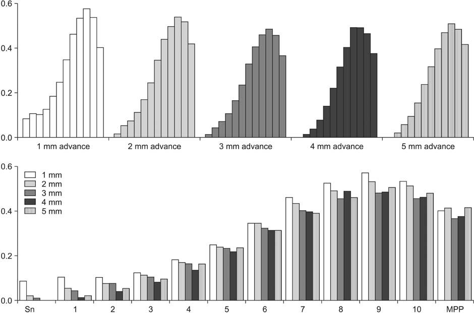
Figure 12 Soft-tissue movement ratio of Curve 2. The curves were divided into 11 sections, and ten equidistant points were added on each curve. Sn, Subnasale; MPP, most protrusive point.

Figure 13 Soft-tissue movement ratio of Curve 3. The curves were divided into 11 sections, and ten equidistant points were added on each curve. LIPP, Left inferior philtrum point; MPP, most protrusive point.
Cited by 2 articles
-
The influence of age on lip-line cant in adults: a cross-sectional study
Sung Hwan Choi, Jung Suk Kim, Cheol Soon Kim, Chung Ju Hwang
Korean J Orthod. 2016;46(2):81-86. doi: 10.4041/kjod.2016.46.2.81.Three-dimensional changes in lip vermilion morphology of adult female patients after extraction and non-extraction orthodontic treatment
Zhi-Yu Liu, Jie Yu, Fan-Fan Dai, Ruo-Ping Jiang, Tian-Min Xu
Korean J Orthod. 2019;49(4):222-234. doi: 10.4041/kjod.2019.49.4.222.
Reference
-
1. Burstone CJ. Lip posture and its significance in treatment planning. Am J Orthod. 1967; 53:262–284.
Article2. Ricketts RM. Esthetics, environment, and the law of lip relation. Am J Orthod. 1968; 54:272–289.
Article3. Tweed CH. The Frankfort-mandibular plane angle in orthodontic diagnosis, classification, treatment planning, and prognosis. Am J Orthod Oral Surg. 1946; 32:175–230.
Article4. Burcal RG, Laskin DM, Sperry TP. Recognition of profile change after simulated orthognathic surgery. J Oral Maxillofac Surg. 1987; 45:666–670.
Article5. Bittner C, Pancherz H. Facial morphology and malocclusions. Am J Orthod Dentofacial Orthop. 1990; 97:308–315.
Article6. McCance AM, Moss JP, Fright WR, James DR, Linney AD. A three-dimensional analysis of bone and soft tissue to bone ratio of movements in 17 Skeletal II patients following orthognathic surgery. Eur J Orthod. 1993; 15:97–106.
Article7. Kim YI, Kim JR, Park SB. Three-dimensional analysis of midfacial soft tissue changes according to maxillary superior movement after horizontal osteotomy of the maxilla. J Craniofac Surg. 2010; 21:1587–1590.
Article8. Solem RC, Marasco R, Guiterrez-Pulido L, Nielsen I, Kim SH, Nelson G. Three-dimensional soft-tissue and hard-tissue changes in the treatment of bimaxillary protrusion. Am J Orthod Dentofacial Orthop. 2013; 144:218–228.
Article9. Kau CH, Hunter LM, Hingston EJ. A different look: 3-dimensional facial imaging of a child with Binder syndrome. Am J Orthod Dentofacial Orthop. 2007; 132:704–709.
Article10. Baik HS, Jeon JM, Lee HJ. Facial soft-tissue analysis of Korean adults with normal occlusion using a 3-dimensional laser scanner. Am J Orthod Dentofacial Orthop. 2007; 131:759–766.
Article11. Mirabella D, Bacconi S, Gracco A, Lombardo L, Siciliani G. Upper lip changes correlated with maxillary incisor movement in 65 orthodontically treated adult patients. World J Orthod. 2008; 9:337–348.12. Ramos AL, Sakima MT, Pinto Ados S, Bowman SJ. Upper lip changes correlated to maxillary incisor retraction--a metallic implant study. Angle Orthod. 2005; 75:499–505.13. Jacobs JD. Vertical lip changes from maxillary incisor retraction. Am J Orthod. 1978; 74:396–404.
Article14. Gwilliam JR, Cunningham SJ, Hutton T. Reproducibility of soft tissue landmarks on three-dimensional facial scans. Eur J Orthod. 2006; 28:408–415.
Article15. Lee WJ, Lee KJ, Yu HS, Baik HS. Lip and perioral soft tissue changes after bracket bonding using 3-D laser scanner. Korean J Orthod. 2011; 41:411–422.
Article16. Wisth J. Soft tissue response to upper incisor retraction in boys. Br J Orthod. 1974; 1:199–204.
Article17. Talass MF, Talass L, Baker RC. Soft-tissue profile changes resulting from retraction of maxillary incisors. Am J Orthod Dentofacial Orthop. 1987; 91:385–394.
Article18. Hershey HG. Incisor tooth retraction and subsequent profile change in postadolescent female patients. Am J Orthod. 1972; 61:45–54.
Article19. Roos N. Soft-tissue profile changes in class II treatment. Am J Orthod. 1977; 72:165–175.
Article
- Full Text Links
-
- Actions
-
Cited
- CITED
-
- Close
- Share
-
- Similar articles
-
- Anterior esthetic restoration using DSD (digital smile design) for a patient with congenital missing tooth of maxillary central incisor
- A Morphometric Study of Primary Anterior Zirconia Crowns in Korean Tooth Models
- Three-dimensional finite element analysis of the phenomenon produced during retraction of four maxillary incisors
- A roentgenocephalometric study of the bony structure and its profile
- Finite element analysis of anterior maxillary segmental distraction osteogenesis using asymmetric distractors in patients with unilateral cleft lip and palate
