Camouflage treatment in adult skeletal Class III cases by extraction of two lower premolars
- Affiliations
-
- 1Department of Orthodontics, School of Stomatology, The Fourth Military Medical University, China. ningfang327@163.com
- KMID: 2273226
- DOI: http://doi.org/10.4041/kjod.2010.40.5.349
Abstract
OBJECTIVE
The purpose of this study was to evaluate the dentoskeletal and soft tissue profile changes after extraction of two lower first or second premolars in "borderline" adult skeletal Class III cases.
METHODS
Twenty-eight patients with "borderline" skeletal Class III malocclusion were studied. All of them were treated by extraction of two lower first or second premolars. Lateral cephalometric radiographs taken at the start and end of treatment were analysed. Twenty-five cephalometric variables were calculated and paired t-tests were performed.
RESULTS
After treatment, no significant changes were noted in the skeletal parameters (p > or = 0.05). Regarding the dental parameters, the L1-MP angle decreased by 8.1degrees, the U1-L1 angle increased by 7.7degrees (p < 0.01), the overjet distance increased by 5.7 mm (p < 0.01), the L1-NB angle decreased by 7.3degrees and the L1-NB distance decreased by 4.8 mm (p < 0.01). The soft tissue parameters of Li-E, Li-H and Li-RL2 distance decreased by 3.2 mm, 3.4 mm and 4.1 mm respectively (p < 0.01).
CONCLUSIONS
Orthodontic camouflage treatment by extraction of two lower first or second premolars provides a viable treatment alternative for "borderline" skeletal Class III cases to achieve a good occlusal relationship.
Figure
-
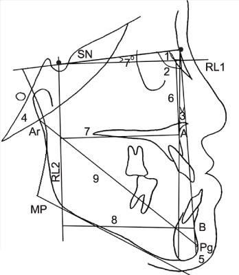
Fig. 1 Skeletal measurements used in the study. SN indicates Sella-nasion plane; RL1, horizontal reference line; RL2, vertical reference line; 1, SNA; 2, SNB; 3, ANB; 4, SN-MP; 5, Pg-NB; 6, A-RL1; 7, A-RL2; 8, B-RL2; 9, Ar-Pg.

Fig. 2 Dental measurements used in the study. SN indicates Sella-nasion plane; RL1, horizontal reference line; RL2, vertical reference line; 1, U1-SN; 2, L1-MP; 3, U1-L1; 4, U1-NA (degree); 5, U1-NA (mm); 6, L1-NB (degree); 7, L1-NB (mm); 8, OP-FH; 9, overjet.

Fig. 3 Soft tissue measurements used in the study. SN indicates Sella-nasion plane; RL1, horizontal reference line; RL2, vertical reference line; 1, upper lip to E plane; 2, lower lip to E plane; 3, lower lip to H line; 4, Cm-Sn-Ls; 5, Ls-RL2; 6, Li-RL2; 7, A-Ls.
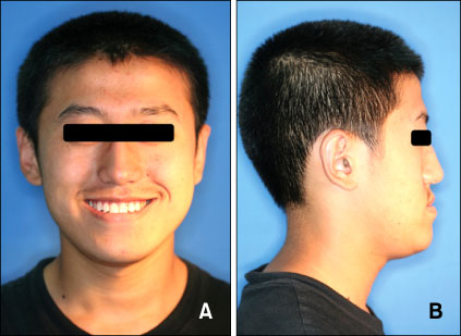
Fig. 4 Pretreatment facial photographs. A, Frontal view; B, lateral view
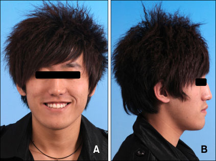
Fig. 5 Posttreatment facial photographs. A, Frontal view; B, lateral view.

Fig. 6 Pretreatment intraoral photographs. A, Lateral view on the right side; B, frontal view; C, lateral view on the left side; D, occlusal view of maxillary dentition; E, occlusal view of mandibular dentition; F, lateral view of anterior teeth.
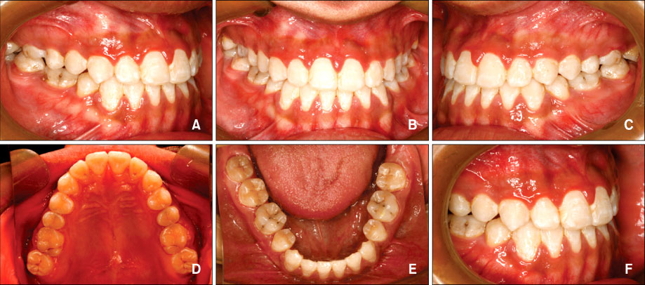
Fig. 7 Posttreatment intraoral photographs. A, Lateral view on the right side; B, frontal view; C, lateral view on the left side; D, occlusal view of maxillary dentition; E, occlusal view of mandibular dentition; F, lateral view of anterior teeth.
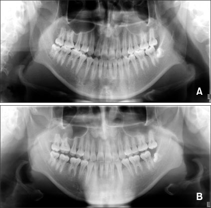
Fig. 8 Panoramic radiographs. A, Pretreatment; B, posttreatment.
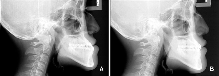
Fig. 9 Lateral cephalometric radiographs. A, Pretreatment; B, posttreatment.

Fig. 10 Superimposition of pretreatment and posttreatment cephalometric tracings.
Cited by 1 articles
-
Comparison of soft tissue changes between incisor tipping and translation after premolar extraction
Wonkyeong Baik, Sung-Hwan Choi, Jung-Yul Cha, Hyung-Seog Yu, Kee-Joon Lee
Korean J Orthod. 2022;52(1):42-52. doi: 10.4041/kjod.2022.52.1.42.
Reference
-
1. Lin J, Gu Y. Preliminary investigation of nonsurgical treatment of severe skeletal Class III malocclusion in the permanent dentition. Angle Orthod. 2003. 73:401–410.2. Guyer EC, Ellis EE 3rd, McNamara JA Jr, Behrents RG. Components of Class III malocclusion in juveniles and adolescents. Angle Orthod. 1986. 56:7–30.3. Dietrich UC. Morphological variability of skeletal Class 3 relationships as revealed by cephalometric analysis. Rep Congr Eur Orthod Soc. 1970. 131–143.4. Duan YZ, Yang ZH, Leng J. Effects of face mask/Class III elastic therapy on severe skeletal crossbite. J Modern Stomatol. 2003. 17:251–252.5. Gu Y, Rabie AB, Hägg U. Treatment effects of simple fixed appliance and reverse headgear in correction of anterior crossbites. Am J Orthod Dentofacial Orthop. 2000. 117:691–699.
Article6. Uner O, Yüksel S, Uçüncü N. Long-term evaluation after chincap treatment. Eur J Orthod. 1995. 17:135–141.
Article7. Cha KS. Skeletal changes of maxillary protraction in patients exhibiting skeletal class III malocclusion: a comparison of three skeletal maturation groups. Angle Orthod. 2003. 73:26–35.8. Tollaro I, Baccetti T, Franchi L. Craniofacial changes induced by early functional treatment of Class III malocclusion. Am J Orthod Dentofacial Orthop. 1996. 109:310–318.
Article9. Lin J, Gu Y. Lower second molar extraction in correction of severe skeletal Class III malocclusion. Angle Orthod. 2006. 76:217–225.10. Sato S. Case report: developmental characterization of skeletal Class III malocclusion. Angle Orthod. 1994. 64:105–111.11. Lew KK. Soft tissue profile changes following orthodontic treatment of Chinese adults with Class III malocclusion. Int J Adult Orthodon Orthognath Surg. 1990. 5:59–65.12. Demir A, Uysal T, Sari Z, Basciftci FA. Effects of camouflage treatment on dentofacial structures in Class II division 1 mandibular retrognathic patients. Eur J Orthod. 2005. 27:524–531.
Article13. Nalbantgil D, Arun T, Sayinsu K, Fulya I. Skeletal, dental and soft-tissue changes induced by the Jasper Jumper appliance in late adolescence. Angle Orthod. 2005. 75:426–436.14. Ning F, Duan Y, Huo N. Camouflage treatment in skeletal Class III cases combined with severe crowding by extraction of four premolars. Orthod Waves. 2009. 68:80–87.
Article15. Rabie AB, Wong RW, Min GU. Treatment in borderline Class III malocclusion: orthodontic camouflage (extraction) versus orthognathic surgery. Open Dent J. 2008. 2:38–48.
Article16. Cassidy DW Jr, Herbosa EG, Rotskoff KS, Johnston LE Jr. A comparison of surgery and orthodontics in "borderline" adults with Class II, division 1 malocclusions. Am J Orthod Dentofacial Orthop. 1993. 104:455–470.
Article17. Kerr WJ, Miller S, Dawber JE. Class III malocclusion: surgery or orthodontics? Br J Orthod. 1992. 19:21–24.
Article18. Zeng XL, Lin JX, Huang JF. Skeletal crossbite: surgery or orthodontics? West China J Stomatol. 1985. 3:233–237.19. Stellzig-Eisenhauer A, Lux CJ, Schuster G. Treatment decision in adult patients with Class III malocclusion: orthodontic therapy or orthognathic surgery? Am J Orthod Dentofacial Orthop. 2002. 122:27–37.
Article20. Pellegrino G. Italian board of orthodontics: case N. 2 adult malocclusion. Prog Orthod. 2005. 6:102–112.21. Fukui T, Tsuruta M. Invisible treatment of a Class III female adult patient with severe crowding and cross-bite. J Orthod. 2002. 29:267–275.
Article
- Full Text Links
-
- Actions
-
Cited
- CITED
-
- Close
- Share
-
- Similar articles
-
- Camouflage treatment by backward rotation of the mandible for a severe skeletal Class III malocclusion with aplastic anemia: A case report
- Lower incisor extraction for dental camouflage
- A study of the calcification of the second and the third molars in skeletal Class II and III malocclusions
- A study on the frequency of tooth extraction for orthodontic treatment
- Morphology of mandibular symphysis and positioning of lower incisors in the skeletal Class III malocclusions
