Correction of palatally displaced maxillary lateral incisors without brackets
- Affiliations
-
- 1Department of Clinical Orthodontics, Graduate School of Clinical Dentistry, Ewha Womans University, Seoul, Korea. yschun@ewha.ac.kr
- KMID: 2272256
- DOI: http://doi.org/10.4041/kjod.2013.43.4.201
Abstract
- This article describes the orthodontic treatment of a 25-year-old Korean female patient with anterior crowding, including palatally displaced lateral incisors. Her facial profile was satisfactory, but 3.5 mm of maxillary anterior crowding was observed. To correct this crowding, we decided to minimize the use of the conventional fixed orthodontic appliances and employed a less bulky and more aesthetic appliance for applying light continuous force. We determined the final positions of the maxillary teeth via a working model for diagnostic set up and achieved space gaining and alignment with simple Ni-Ti spring and stainless steel round tubes. Tooth alignment was achieved efficiently and aesthetically without the conventional brackets.
Keyword
MeSH Terms
Figure
-
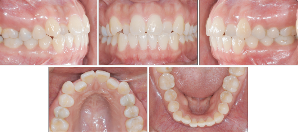
Figure 1 Pretreatment intraoral photographs.
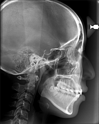
Figure 2 Pretreatment lateral cephalograph.
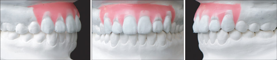
Figure 3 Working model for diagnostic set up.
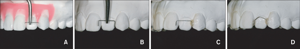
Figure 4 Fabrication of Ni-Ti spring. A, The necessary space was determined in the working model. B, A 0.012-inch nickel-titanium (Ni-Ti) wire was bent to create a Ni-Ti spring. C, D, Orthodontic adhesive resin was used to attach this spring to the canine and central incisor on each side.
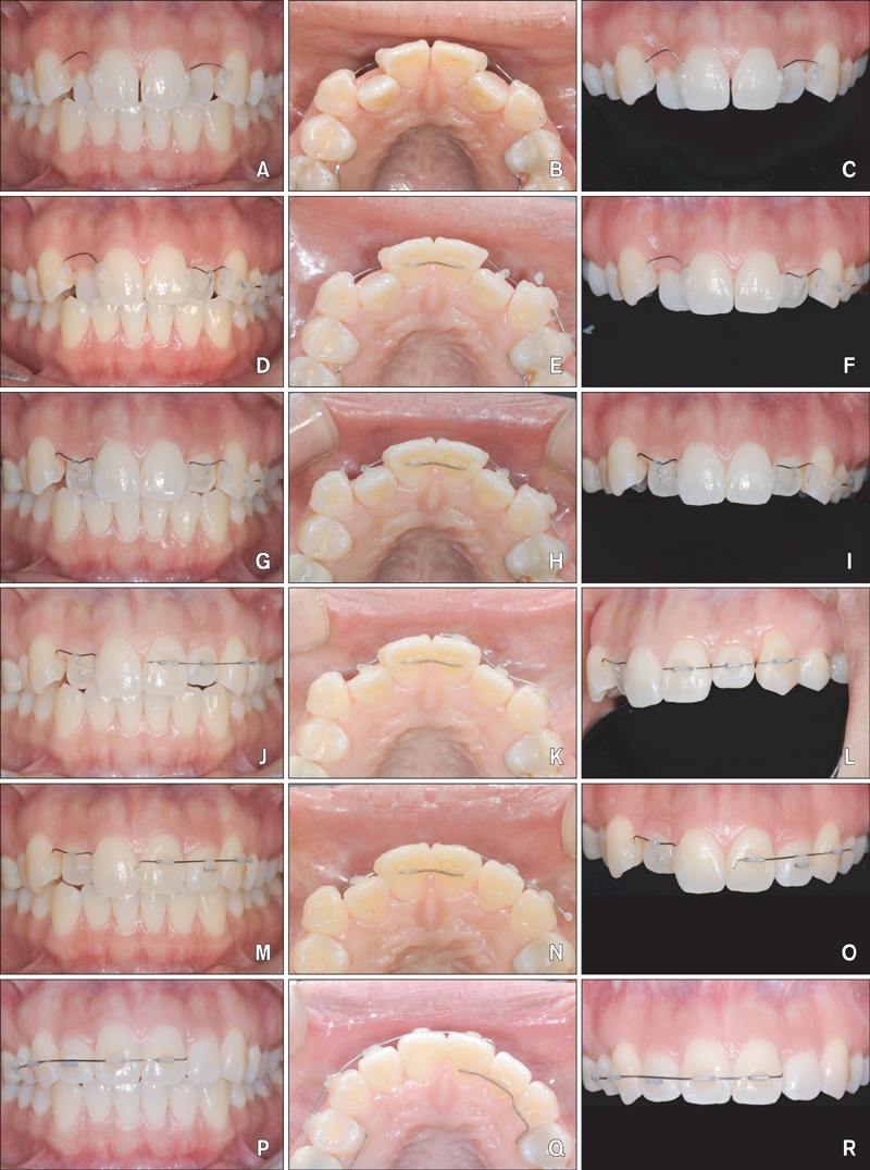
Figure 5 Treatment progress. A-C, 0.012-inch nickel-titanium (Ni-Ti) wires were bent to create Ni-Ti springs that were bonded proximally to the canines and central incisors by using orthodontic adhesive resin. Stripping of the central incisors was performed before the attachment. D-F, After the adequate space was regained, a 0.008-inch ligature wire was attached to the surface of the left lateral incisor and ligated to the Ni-Ti wire for intrusion; the left canine and premolars were connected to serve as the anchor. G-I, Intrusion of the left lateral incisor was achieved and intrusion of the right lateral incisor was initiated by using the same method. J-L, After the left lateral incisor intrusion, alignment was initiated by using round tubes and a 0.012-inch Ni-Ti wire. M-O, Leveling was performed by wire overlay above the lateral incisor tubes. P-R, A lingual retainer was attached to the left anterior region and the right-side alignment was performed by using the same method.
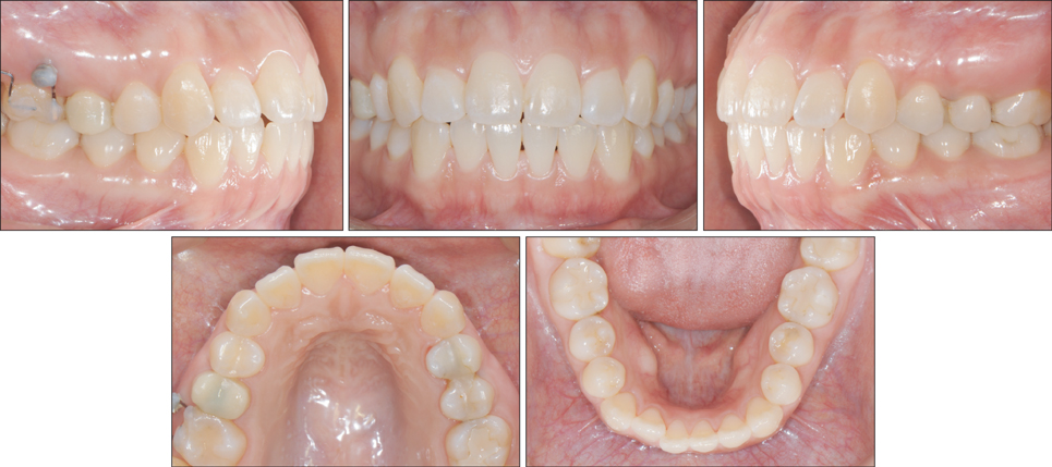
Figure 6 Post-treatment intraoral photographs.
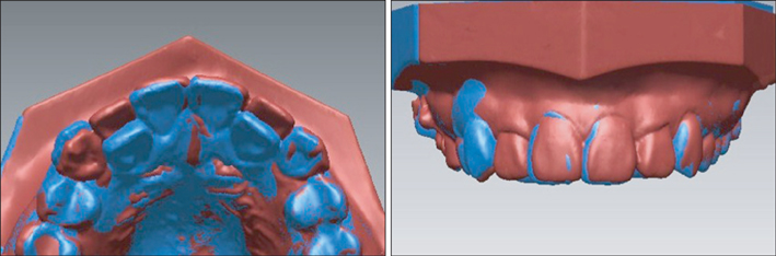
Figure 7 Model superimposition. Alignment was achieved by moving both the lateral incisors labially by 3.0 - 3.5 mm and the right canine palatally by approximately 1 mm. The midline was shifted approximately 1 mm to the left.

Figure 8 Cone-beam computed tomography superimposition of the left maxillary lateral incisor. The images show that the tooth axis was upright at the initial assessment, but the correct amount of torque developed with the appropriate uncontrolled tipping.
Cited by 2 articles
-
Maxillary molar derotation and distalization by using a nickel-titanium wire fabricated on a setup model
Jong Moon Jung, Young Joo Wi, Hyun Mo Koo, Min Ji Kim, Youn Sic Chun
Korean J Orthod. 2017;47(4):268-274. doi: 10.4041/kjod.2017.47.4.268.Treatment of Class I crowding using simple tubes bonded with customized resin coverings: A case report
Seo-Rin Jeong, Hye-In Kim, Sung-Hoon Lim
Korean J Orthod. 2019;49(2):116-123. doi: 10.4041/kjod.2019.49.2.116.
Reference
-
1. Ponitz RJ. Invisible retainers. Am J Orthod. 1971; 59:266–272.
Article2. Sheridan JJ, LeDoux W, McMinn R. Essix retainers: fabrication and supervision for permanent retention. J Clin Orthod. 1993; 27:37–45.3. Phan X, Ling PH. Clinical limitations of Invisalign. J Can Dent Assoc. 2007; 73:263–266.4. Proffit WP, Fields HW, Sarver DM. Contemporary orthodontics. 4th ed. St. Louis: Mosby-Year Book Inc;2007. p. 559–560.5. Park SH, Lee YK, Chun YS. Correction of palatally displaced maxillary lateral incisors using a tube system. J Clin Orthod. 2008; 42:461–465.
- Full Text Links
-
- Actions
-
Cited
- CITED
-
- Close
- Share
-
- Similar articles
-
- Clinical effects of different prescriptions on the inclination of maxillary and mandibular incisors by using passive self-ligating brackets
- Color Distribution of Maxillary Permanent Incisors in Korean Pediatric Patients Using a Spectrophotometer
- Color Comparison of Maxillary Primary Anterior Teeth and Various Composite Resins using a Spectrophotometer
- The positioning errors in bonding lingual brackets
- Management of Displaced Maxillary Canines by Extraction of the Primary Canine: Factors Affecting Treatment Outcome
