Korean J Orthod.
2013 Aug;43(4):178-185. 10.4041/kjod.2013.43.4.178.
Soft-tissue thickness of South Korean adults with normal facial profiles
- Affiliations
-
- 1Department of Orthodontics, School of Dentistry, Dankook University, Cheonan, Korea. kscha@dankook.ac.kr
- KMID: 2272253
- DOI: http://doi.org/10.4041/kjod.2013.43.4.178
Abstract
OBJECTIVE
To standardize the facial soft-tissue characteristics of South Korean adults according to gender by measuring the soft-tissue thickness of young men and women with normal facial profiles by using three-dimensional (3D) reconstructed models.
METHODS
Computed tomographic images of 22 men aged 20 - 27 years and 18 women aged 20 - 26 years with normal facial profiles were obtained. The hard and soft tissues were three-dimensionally reconstructed by using Mimics software. The soft-tissue thickness was measured from the underlying bony surface at bilateral (frontal eminence, supraorbital, suborbital, inferior malar, lateral orbit, zygomatic arch, supraglenoid, gonion, supraM2, occlusal line, and subM2) and midline (supraglabella, glabella, nasion, rhinion, mid-philtrum, supradentale, infradentale, supramentale, mental eminence, and menton) landmarks.
RESULTS
The men showed significantly thicker soft tissue at the supraglabella, nasion, rhinion, mid-philtrum, supradentale, and supraglenoid points. In the women, the soft tissue was significantly thicker at the lateral orbit, inferior malar, and gonion points.
CONCLUSIONS
The soft-tissue thickness in different facial areas varies according to gender. Orthodontists should use a different therapeutic approach for each gender.
Keyword
Figure
-

Figure 1 Method used for the three-demensional computed tomography-image reconstruction.
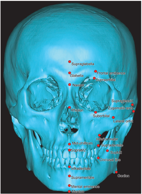
Figure 2 Landmarks on the three-demensional reconstructed hard-tissue model.
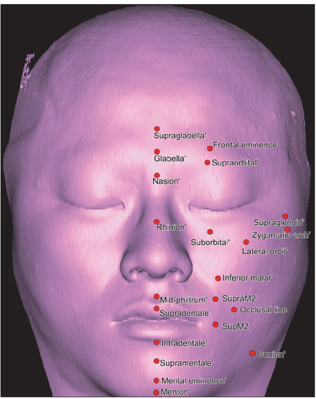
Figure 3 Landmarks on the three-dimensional reconstructed soft-tissue model.
Reference
-
1. Ackerman JL, Proffit WR, Sarver DM. The emerging soft tissue paradigm in orthodontic diagnosis and treatment planning. Clin Orthod Res. 1999; 2:49–52.
Article2. Simpson E, Henneberg M. Variation in soft-tissue thicknesses on the human face and their relation to craniometric dimensions. Am J Phys Anthropol. 2002; 118:121–133.
Article3. Kim HJ, Kang MK, Hu KS, Kim CH, Chung IH. Measurements of the Korean facial thickness. Korean J Leg Med. 1999; 23:117–121.4. Baek SH, Yang WS. A soft tissue analysis on facial esthetics of Korean young adults. Korean J Orthod. 1991; 21:131–170.5. Kang SG, Lee YJ, Park YG. A comparative study of soft tissue profile between Korean and Caucasian young adults under NHP. Korean J Orthod. 2003; 33:323–337.6. Jeong HG, Kim KD, Han SH, Shin DW, Hu KS, Lee JB, et al. Measurement of facial soft tissues thickness using 3D computed tomographic images. Korean J Oral Maxillofac Radiol. 2006; 36:49–54.7. Weisell RC. Body mass index as an indicator of obesity. Asia Pac J Clin Nutr. 2002; 11:Suppl 8. S681–S684.
Article8. Lundström A, Forsberg CM, Peck S, McWilliam J. A proportional analysis of the soft tissue facial profile in young adults with normal occlusion. Angle Orthod. 1992; 62:127–133.9. Lee JH, Nahm DS. A proportional analysis of soft tissue profile in korean young adults. Korean J Orthod. 1994; 24:405–418.10. White SC. Cone-beam imaging in dentistry. Health Phys. 2008; 95:628–637.
Article11. Swennen GR, Schutyser F. Three-dimensional cephalometry: spiral multi-slice vs cone-beam computed tomography. Am J Orthod Dentofacial Orthop. 2006; 130:410–416.
Article12. Coben SE. The integration of facial skeletal variations. Am J Orthod. 1955; 41:407–434.13. Kim KD, Ruprecht A, Wang G, Lee JB, Dawson DV, Vannier MW. Accuracy of facial soft tissue thickness measurements in personal computer-based multiplanar reconstructed computed tomographic images. Forensic Sci Int. 2005; 155:28–34.
Article14. Phillips VM, Smuts NA. Facial reconstruction: utilization of computerized tomography to measure facial tissue thickness in a mixed racial population. Forensic Sci Int. 1996; 83:51–59.
Article15. Aulsebrook WA, Becker PJ, Işcan MY. Facial soft-tissue thicknesses in the adult male Zulu. Forensic Sci Int. 1996; 79:83–102.
Article16. Hashim HA, AlBarakati SF. Cephalometric soft tissue profile analysis between two different ethnic groups: a comparative study. J Contemp Dent Pract. 2003; 4:60–73.
Article17. Sahni D, Sanjeev , Singh G, Jit I, Singh P. Facial soft tissue thickness in northwest Indian adults. Forensic Sci Int. 2008; 176:137–146.
Article18. Codinha S. Facial soft tissue thicknesses for the Portuguese adult population. Forensic Sci Int. 2009; 184:80.e1–80.e7.
Article19. Domaracki M, Stephan CN. Facial soft tissue thicknesses in Australian adult cadavers. J Forensic Sci. 2006; 51:5–10.
Article20. Kohn LA, Cheverud JM, Bhatia G, Commean P, Smith K, Vannier MW. Anthropometric optical surface imaging system repeatability, precision, and validation. Ann Plast Surg. 1995; 34:362–371.
Article
- Full Text Links
-
- Actions
-
Cited
- CITED
-
- Close
- Share
-
- Similar articles
-
- A longitudinal study on growth changes of soft tissue profiles in Koreans with normal occlusion
- A longitudinal study of soft-tissue facial profile changes in Korean children
- A longitudinal study on the soft tissue facial profile change: Study report from 9 to 13 years of age
- A roentgenocephalometric study of korean soft tissue profile
- Evaluation of facial soft tissue thickness in asymmetric mandibular deformities after orthognathic surgery
