Usefulness of the Computed Tomography Venography for Evaluation of Leg Edema Including Deep Vein Thrombosis in Rehabilitation Patients
- Affiliations
-
- 1Department of Physical Medicine & Rehabilitation, Dongguk University College of Medicine, Goyang, Korea. hjrhee1@dumc.or.kr
- 2Department of Radiology, Dongguk University College of Medicine, Goyang, Korea.
- KMID: 2267092
- DOI: http://doi.org/10.5535/arm.2014.38.6.812
Abstract
OBJECTIVE
To investigate the usefulness of computed tomography venography (CTV) for evaluation of leg swelling, especially deep vein thrombosis (DVT), in rehabilitation patients.
METHODS
A hundred twenty-three patients, who had performed CTV performed because of suspected DVT in our clinic, were enrolled. We performed chart reviews retrospectively and categorized CTV findings as follows: DVT distal to inguinal ligament and no compression lesion; DVT proximal to inguinal ligament and no compression lesion; DVT distal to inguinal ligament and anatomical variant (for example, May-Thurner syndrome); DVT due to compression of mass (cancer or cyst); DVT and other incidental abnormal finding; and no DVT and other possible causes of leg swelling.
RESULTS
DVTs were found in 65 (53%) patients. DVTs were found at distal level (thigh or lower leg) to inguinal ligament in 47 patients. DVTs were found at proximal to inguinal ligament, usually undetectable with duplex ultrasonography, in 6 patients. DVTs caused by external compression, such as femoral vein and cancer mass, were found in 12 patients (10%), which are also not easily detected with duplex ultrasonography. Other various causes of leg edema without DVT were found in 22 (18%) patients.
CONCLUSION
CTV can evaluate more extensively venous problems in the pelvis and abdomen and detect other possible causes of leg swelling. Therefore, CTV can be a useful tool not only for easy detection of DVT but also for evaluating differential diagnosis of leg edema in rehabilitation patients.
Keyword
MeSH Terms
Figure
-
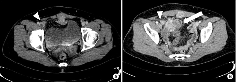
Fig. 1 The computed tomography venography reveals low signal intensity lesion, which means deep vein thrombosis (DVT), inside of right iliac vein (arrowhead) (A) and the well enhancing mass lesions at right lower abdomen (white arrow) compress right external iliac vein (arrowhead) proximal to DVT lesion (B).
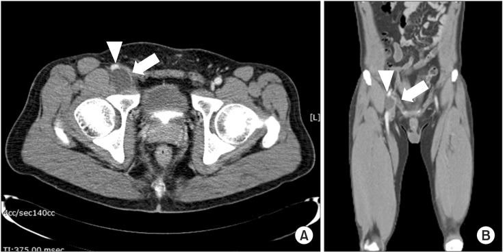
Fig. 2 This image shows low density 2.8×2.9×2.1 cm sized mass lesion (white arrow) in posterolateral area to right common femoral vein (arrowhead) in axial view (A) and the mass (white arrow) compresses right common femoral vein (arrowhead) in coronal view (B).
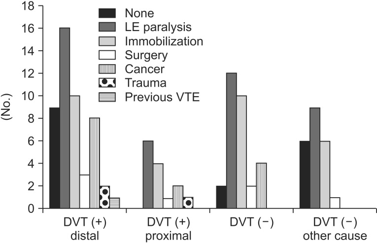
Fig. 3 The graph shows distributions of risk factors of VTE among four groups: DVT (+) distal, DVT distal to inguinal ligament; DVT (+) proximal, DVT proximal to inguinal ligament (in abdomen/pelvis) or with compression of other structures; DVT (-), no DVT detected; and DVT (-) other causes, no DVT and other causes of leg swelling detected). The relative proportion of LE paralysis in DVT (-) and DVT (-) with other cause groups was higher, similar to DVT (+) group. Therefore in the patients with LE paralysis usually present in the department of rehabilitation there are various causes of leg swelling and some causes can be found more easily by computed tomography venography than other study. DTV, deep vein thrombosis; VTE, venous thromboembolism; LE, lower extremity.
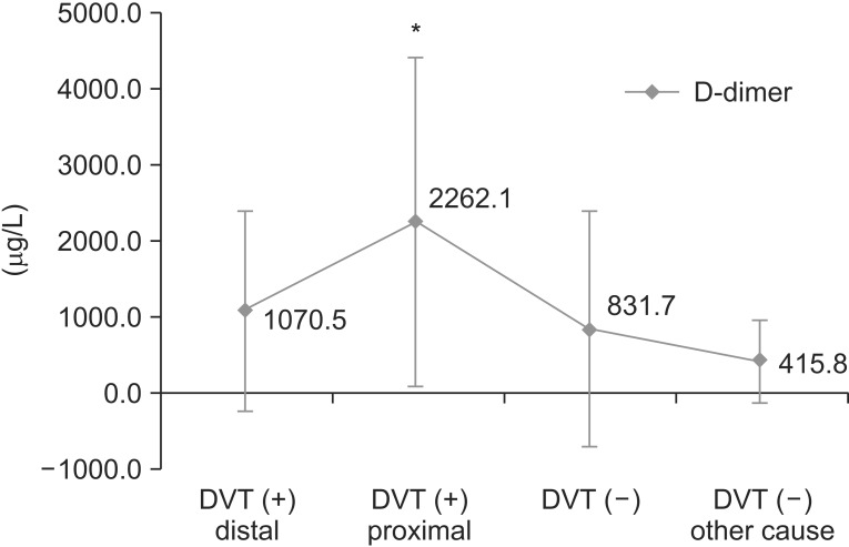
Fig. 4 This graph shows the D-dimer values of four groups. D-dimer values of deep vein thrombosis (DVT) proximal group (groups 2, 3, and 4) were significantly different from those of the other three groups. *p<0.05, result from ANOVA with post-hoc analysis.
Cited by 1 articles
-
Leg Swelling Caused by Heterotopic Ossification Mimicking Deep Vein Thrombosis in a Paraplegic Patient
Jin Hyuk Bang, Keun-Tae Cho, Ho Jun Lee
Korean J Neurotrauma. 2015;11(2):158-161. doi: 10.13004/kjnt.2015.11.2.158.
Reference
-
1. Ely JW, Osheroff JA, Chambliss ML, Ebell MH. Approach to leg edema of unclear etiology. J Am Board Fam Med. 2006; 19:148–160. PMID: 16513903.
Article2. Warlow C, Ogston D, Douglas AS. Deep venous thrombosis of the legs after stroke. Part 2: Natural history. Br Med J. 1976; 1:1181–1183. PMID: 1268615.3. Kelly J, Rudd A, Lewis R, Hunt BJ. Venous thromboembolism after acute stroke. Stroke. 2001; 32:262–267. PMID: 11136946.
Article4. Byrne JJ. Phlebitis; a study of 748 cases at the Boston City Hospital. N Engl J Med. 1955; 253:579–586. PMID: 13265999.5. Collins R, Scrimgeour A, Yusuf S, Peto R. Reduction in fatal pulmonary embolism and venous thrombosis by perioperative administration of subcutaneous heparin: overview of results of randomized trials in general, orthopedic, and urologic surgery. N Engl J Med. 1988; 318:1162–1173. PMID: 3283548.
Article6. Wijdicks EF, Scott JP. Pulmonary embolism associated with acute stroke. Mayo Clin Proc. 1997; 72:297–300. PMID: 9121173.7. Bromfield EB, Reding MJ. Relative risk of deep venous thrombosis or pulmonary embolism post-stroke based on ambulatory status. Neurorehabil Neural Repair. 1988; 2:51–57.
Article8. Ko HY, Shin YB, Jho SK. Incidence of deep vein thrombosis in spinal cord injury. J Korean Acad Rehabil Med. 2005; 29:359–364.9. Han TR, Lim SJ, Lee HJ. Deep vein thrombosis in rehabilitation inpatients. J Korean Acad Rehabil Med. 2001; 25:827–835.10. Anderson FA Jr, Wheeler HB, Goldberg RJ, Hosmer DW, Patwardhan NA, Jovanovic B, et al. A populationbased perspective of the hospital incidence and casefatality rates of deep vein thrombosis and pulmonary embolism: the Worcester DVT Study. Arch Intern Med. 1991; 151:933–938. PMID: 2025141.
Article11. Lim KE, Hsu WC, Hsu YY, Chu PH, Ng CJ. Deep venous thrombosis: comparison of indirect multidetector CT venography and sonography of lower extremities in 26 patients. Clin Imaging. 2004; 28:439–444. PMID: 15531146.12. Byun SS, Kim JH, Kim YJ, Jeon YS, Park CH, Kim WH. Evaluation of deep vein thrombosis with multidetector row CT after orthopedic arthroplasty: a prospective study for comparison with Doppler sonography. Korean J Radiol. 2008; 9:59–66. PMID: 18253077.
Article13. Goodman LR, Stein PD, Matta F, Sostman HD, Wakefield TW, Woodard PK, et al. CT venography and compression sonography are diagnostically equivalent : data from PIOPED II. AJR Am J Roentgenol. 2007; 189:1071–1076. PMID: 17954642.14. Loud PA, Katz DS, Klippenstein DL, Shah RD, Grossman ZD. Combined CT venography and pulmonary angiography in suspected thromboembolic disease: diagnostic accuracy for deep venous evaluation. AJR Am J Roentgenol. 2000; 174:61–65. PMID: 10628455.15. Geerts WH, Bergqvist D, Pineo GF, Heit JA, Samama CM, Lassen MR, et al. Prevention of venous thromboembolism: American College of Chest Physicians Evidence-Based Clinical Practice Guidelines (8th Edition). Chest. 2008; 133(6 Suppl):381S–453S. PMID: 18574271.16. Cham MD, Yankelevitz DF, Shaham D, Shah AA, Sherman L, Lewis A, et al. Deep venous thrombosis: detection by using indirect CT venography. The Pulmonary Angiography-Indirect CT Venography Cooperative Group. Radiology. 2000; 216:744–751. PMID: 10966705.17. Knapton S, Pandian G. Lower limb peripheral vascular disease. In : Braddom RL, editor. Physical medicine and rehabilitation. 4th ed. Philadelphia: Saunders;2011. p. 1347–1370.18. Ley EJ, Hood DB, Leke MA, Rao RK, Rowe VL, Weaver FA. Endovascular management of iliac vein occlusive disease. Ann Vasc Surg. 2004; 18:228–233. PMID: 15253261.
Article19. Chung JW, Yoon CJ, Jung SI, Kim HC, Lee W, Kim YI, et al. Acute iliofemoral deep vein thrombosis: evaluation of underlying anatomic abnormalities by spiral CT venography. J Vasc Interv Radiol. 2004; 15:249–256. PMID: 15028809.
Article20. Bryce TN, Ragnarsson KT, Stein AB, Biering-Sorensen F. Spinal cord injury. In : Braddom RL, editor. Physical medicine and rehabilitation. 4th ed. Philadelphia: Saunders;2011. p. 1293–1346.21. Hou H, Ge Z, Ying P, Dai J, Shi D, Xu Z, et al. Biomarkers of deep venous thrombosis. J Thromb Thrombolysis. 2012; 34:335–346. PMID: 22528325.
Article22. Akman MN, Cetin N, Bayramoglu M, Isiklar I, Kilinc S. Value of the D-dimer test in diagnosing deep vein thrombosis in rehabilitation inpatients. Arch Phys Med Rehabil. 2004; 85:1091–1094. PMID: 15241755.
Article23. Harvey RL, Roth EJ, Yarnold PR, Durham JR, Green D. Deep vein thrombosis in stroke: the use of plasma D-dimer level as a screening test in the rehabilitation setting. Stroke. 1996; 27:1516–1520. PMID: 8784122.
- Full Text Links
-
- Actions
-
Cited
- CITED
-
- Close
- Share
-
- Similar articles
-
- The Incidence of Deep Vein Thrombosis in the Lower Extremity
- Significance of Contrast Enhanced Rapid MR Sequence(True FISP) in Deep Vein Thrombosis
- Clinical Course of Advanced Cancer Patients with Lower-Extremity Edema and Elevated D-dimer Levels who Underwent Computed Tomography Venography
- Gadolinium-Enhanced 3D Fast Imaging Steady-State Procession(FISP) MR Venography in the Deep Vein Thrombosis of Low Extremities
- Incidence of Venous Thromboembolism Using 64 Channel Multidetector Row Computed Tomography-Indirect Venography and Anti-Coagulation Therapy after Total Knee Arthroplasty in Korea
