Ann Rehabil Med.
2012 Apr;36(2):297-302. 10.5535/arm.2012.36.2.297.
Spinal Cord Infarction Caused by Non-dissected and Unruptured Thoracoabdominal Aortic Aneurysm with Intraluminal Thrombus
- Affiliations
-
- 1Department of Rehabilitation Medicine, Chungbuk National University College of Medicine, Cheongju 361-711, Korea. bang@chungbuk.ac.kr
- KMID: 2266773
- DOI: http://doi.org/10.5535/arm.2012.36.2.297
Abstract
- Spinal cord infarction, especially anterior spinal artery syndrome, is a relatively rare disease. We report a case of spinal cord infarction caused by thoracoabdominal aortic aneurysm with intraluminal thrombus. A 52-year-old man presented with sudden onset paraplegia. At first, he was diagnosed with cervical myelopathy due to a C6-7 herniated intervertebral disc, and had an operation for C6-7 discetomy and anterior interbody fusion. Approximately 1 month after the operation, he was transferred to the department of rehabilitation in our hospital. Thoracoabdominal aortic aneurysm with intraluminal thrombus was found incidentally on an enhanced computed tomography scan, and high signal intensities were detected at the anterior horns of gray matter from the T8 to cauda equina level on T2-weighted magnetic resonance imaging. There was no evidence of aortic rupture, dissection, or complete occlusion of the aorta. We diagnosed his case as a spinal cord infarction caused by thoracoabdominal aortic aneurysm with intraluminal thrombus.
Keyword
MeSH Terms
Figure
-
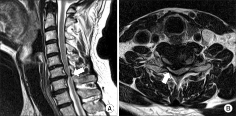
Fig. 1 Magnetic resonance imaging of cervical spine. Sagittal T2-weighted image reveals degeneration and diffuse bulging of disc (arrow) at the C6-7 level (A). Axial T2-weighted image shows a right-side central focal protruded disc (arrow) at the C6-7 level (B).
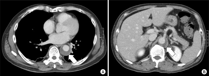
Fig. 2 Enhanced computed tomography scan shows aneurysmal dilatation with intraluminal thrombus (arrow) at the T9 (A) and L1 (B) level.

Fig. 3 Magnetic resonance imaging of thoracolumbar spine. Sagittal T2-weighted image reveals increased intramedullary signal intensity (arrow) from the T8 to cauda equina level (A). Axial T2-weighted image at the T11 level shows bilateral hyperintensities corresponding to the anterior horns of gray matter ('snake eyes' appearance) (arrow) (B).
Reference
-
1. Roppr AH, Samuels MA. Adams and Victor's principles of neurology. 2009. 9th ed. New York: McGraw-Hill Professional;p. 1202–1205.2. Kim HT, Cho WH, Kim HC. Spinal cord ischemia related to infrarenal aortic pathology and surgical procedure. J Korean Soc Vasc Surg. 1999; 15:88–93.3. Oh EJ, Jeong SW, Park JK, Hong KS. Painless aortic dissection simulating Guillain-Barre syndrome. J Korean Soc Clin Neurophysiol. 2005; 7:49–51.4. Han HS, Shin DS, Lee SH, Park SW, Park HY, Chang H, Kim YS, Cho KH, Jang SJ. A case of spinal cord ischemia induced by aortic intramural hematoma. Korean J Stroke. 2004; 6:121–123.5. Weidauer S, Nichtweiss M, Lanfermann H, Zanella FE. Spinal cord infarction: MR imaging and clinical features in 16 cases. Neuroradiology. 2002; 44:851–857. PMID: 12389137.
Article6. Sliwa JA, Maclean IC. Ischemic myelopathy: a review of spinal vasculature and related clinical syndromes. Arch Phys Med Rehabil. 1992; 73:365–372. PMID: 1554311.
Article7. Liu C, Huang Y, Cai HX, Fan SW. Nontraumatic acute paraplegia associated with cervical disk herniation. J Spinal Cord Med. 2010; 33:420–424. PMID: 21061902.
Article8. Cheshire WP, Santos CC, Massey EW, Howard JF Jr. Spinal cord infarction: etiology and outcome. Neurology. 1996; 47:321–330. PMID: 8757000.
- Full Text Links
-
- Actions
-
Cited
- CITED
-
- Close
- Share
-
- Similar articles
-
- Crawford Type III and IV Thoracoabdominal Aortic Aneurysm: 4 Cases Report
- A Case of Paraplegia Following Endovascular Stent Repair of Descending Thoracic Aortic Aneurysm
- Visceral Debranching Thoracic Endovascular Aneurysm Repair for Chronic Dissecting Thoracoabdominal Aortic Aneurysm
- The Clinical Experience of The Descending Thoracic and Thoracoabdominal Aortic Surgery
- Giant Internal Carotid Artery Aneurysm:Posterior Cerebral Artery Territory Infarction After Ligation of the Right Internal Carotid Artery: Case Report
