Effect of the Position of Immobilization Upon the Tensile Properties in Injured Achilles Tendon of Rat
- Affiliations
-
- 1Department of Physical Medicine and Rehabilitation, Institute for Medical Sciences, Chonbuk National University Medical School and Research Institute of Clinical Medicine, Chonbuk National University Hospital, Jeonju, Korea. vivaseo@jbnu.ac.kr
- 2Department of Pharmacology, Institute for Medical Sciences, Chonbuk National University Medical School and Research Institute of Clinical Medicine, Chonbuk National University Hospital, Jeonju, Korea.
- 3Department of Dental Biomaterials, Chonbuk National University School of Dentistry, Jeonju, Korea.
- KMID: 2266644
- DOI: http://doi.org/10.5535/arm.2013.37.1.1
Abstract
OBJECTIVE
To examine the effect of the posture of immobilization upon the tensile properties in injured Achilles tendon of rat for an initial period of immobilization.
METHODS
Forty-two Sprague-Dawley rats were used in the present study. Eighteen rats received a total tenotomy of the right Achilles tendon to mimic total rupture and were divided into three groups comprising of 6 rats each. Ankles of group A were immobilized at 60degrees of plantarflexion. Ankles of group B were immobilized at neutral position. Whereas, those of group C were immobilized at 60degrees of dorsiflexion. Other 18 rats received hemitenotomy to mimic partial rupture and were divided into three groups. The remaining 6 rats were kept free as control. After 14 days, we dissected the tendons and analyzed maximum force, stiffness, and energy uptake during pulling of the tendons until they ruptured. The tendons of 6 rats in each group and control were reserved for histology. Picrosirius staining was done for the analysis of collagen organization.
RESULTS
In total tenotomy, tensile properties were significantly different between the control and the intervention groups (p<0.05). Group C showed relatively higher values than the groups A and B with respect to tensile properties (p>0.05). In partial tenotomy, tensile properties were significantly different between the control and the intervention groups (p<0.05). Group C showed significantly higher value than other intervention groups in terms of maximum force and energy uptake (p<0.05). The semiquantitative histologic grading scores were assigned for collagen organization. The scores for dorsiflexion posture were higher than the ones for plantarflexion.
CONCLUSION
Dorsiflexion posture in partial ruptured Achilles tendon showed better functional recovery than other immobilized postures. In total ruptured case, the tensile properties showed increasing tendency in dorsiflexion posture.
Keyword
MeSH Terms
Figure
-
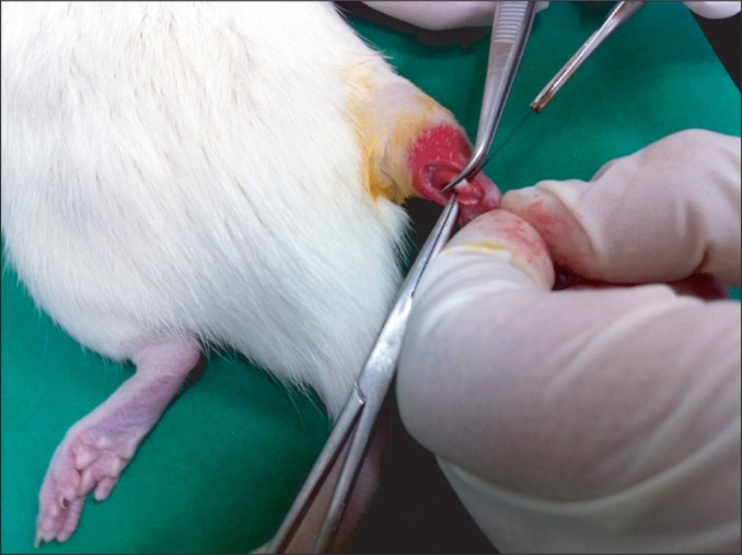
Fig. 1 The figure shows Achilles tendon tenotomy. A 1-cm longitudinal midline incision was made in the skin overlying the Achilles tendon and 1 cm proximal to the calcaneal insertion, and the tendon was separated from the surrounding tissue. Partial or total tenotomy was done at 1 cm proximal to the calcaneal insertion.
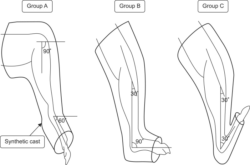
Fig. 2 Schematic diagram of the immobilization. Group A was immobilized at plantarflexion posture of the ankle by 60° with synthetic cast. Group B was immobilized at neutral posture with cast. Group C was immobilized at dorsiflexion posture of the ankle by 60°.
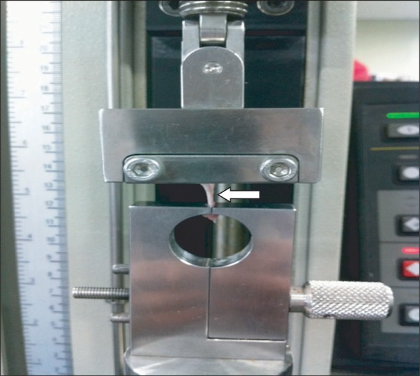
Fig. 3 The figure represents the biomechanical testing machine, Instron. Achilles tendon (arrow) was securely held with clamps and the lesion site was midway between the clamps.
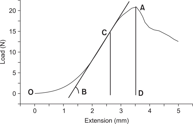
Fig. 4 The results of biomechanical data by Instron. X-axis represents tendon extension in mm, and y-axis represents load in Newton. Tendon is gradually elongated with tension until rupture. Mark A on the graph signifies the maximum force level the tendon can tolerate, in other words, the maximum force at rupture. Mark B signifies the slope of the graph, which represents the stiffness (N/mm), in other words, it means the elasticity of the tendon. Mark C signifies the maximum elastic force applied on the tendon when its elasticity is at the highest. The area bordered by O, A, and D implies the amount of absorbed energy until the tendon rupture.
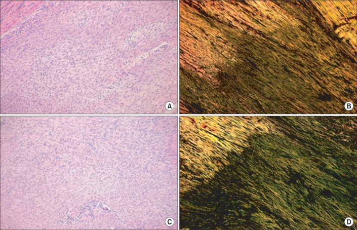
Fig. 5 (A, C) are hematoxylin-eosin (×100) staining and (C, D) are the Picrosirius (×100) staining of longitudinal sections through the rupture site of the Achilles tendon in total tenotomy. Among the total tenotomy rat tendons, (A, B) are from the plantarflexion and (C, D) are from the dorsiflexion posture of immobilization. Green fibers indicate type III collagen in (B, D). In (B, D), the alignment of tendon fibers can be seen. However, (D) of dorsiflexion posture shows denser and well aligned collagen fibers than (B).
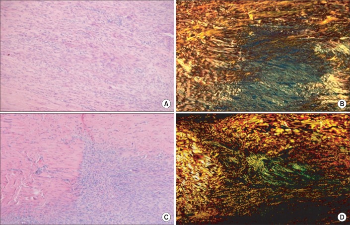
Fig. 6 (A, C) are hematoxylin-eosin (×100) staining and (B, D) are the Picrosirius (×100) staining of longitudinal sections through the rupture site of the Achilles tendon in hemitenotomy. (A, B) are from the plantarflexion and (C, D) are from the dorsiflexion posture of immobilization. Green fibers indicate type III collagen. In hemitenotomy, (D) from dorsiflexion posture also shows denser collagen fibers than (B).
Reference
-
1. Thompson J, Baravarian B. Acute and chronic Achilles tendon ruptures in athletes. Clin Podiatr Med Surg. 2011; 28:117–135. PMID: 21276522.
Article2. Matsumoto F, Trudel G, Uhthoff HK, Backman DS. Mechanical effects of immobilization on the Achilles' tendon. Arch Phys Med Rehabil. 2003; 84:662–667. PMID: 12736878.
Article3. Lee JH, Kim HS, Ahn KH. Effects of local corticosteroid injection and weight bearing on injured Achilles tendon in a rat model. J Korean Acad Rehabil Med. 2002; 26:215–222.4. Longo UG, Ronga M, Maffulli N. Acute ruptures of the achilles tendon. Sports Med Arthrosc. 2009; 17:127–138. PMID: 19440140.
Article5. Amiel D, Woo SL, Harwood FL, Akeson WH. The effect of immobilization on collagen turnover in connective tissue: a biochemical-biomechanical correlation. Acta Orthop Scand. 1982; 53:325–332. PMID: 7090757.
Article6. Harwood FL, Amiel D. Differential metabolic responses of periarticular ligaments and tendon to joint immobilization. J Appl Physiol. 1992; 72:1687–1691. PMID: 1601773.
Article7. Savolainen J, Myllyla V, Myllyla R, Vihko V, Vaananen K, Takala TE. Effects of denervation and immobilization on collagen synthesis in rat skeletal muscle and tendon. Am J Physiol. 1988; 254(6 Pt 2):R897–R902. PMID: 2837917.
Article8. Walsh S, Frank C, Hart D. Immobilization alters cell metabolism in an immature ligament. Clin Orthop Relat Res. 1992; (277):277–288. PMID: 1555352.
Article9. Woo SL, Gomez MA, Sites TJ, Newton PO, Orlando CA, Akeson WH. The biomechanical and morphological changes in the medial collateral ligament of the rabbit after immobilization and remobilization. J Bone Joint Surg Am. 1987; 69:1200–1211. PMID: 3667649.
Article10. Almeida-Silveira MI, Lambertz D, Perot C, Goubel F. Changes in stiffness induced by hindlimb suspension in rat Achilles tendon. Eur J Appl Physiol. 2000; 81:252–257. PMID: 10638386.11. Loitz BJ, Zernicke RF, Vailas AC, Kody MH, Meals RA. Effects of short-term immobilization versus continuous passive motion on the biomechanical and biochemical properties of the rabbit tendon. Clin Orthop Relat Res. 1989; (244):265–271. PMID: 2743669.
Article12. Mullner T, Kwasny O, Reihsner R, Lohnert V, Schabus R. Mechanical properties of a rat patellar tendon stress-shielded in situ. Arch Orthop Trauma Surg. 2000; 120:70–74. PMID: 10653108.
Article13. Trebacz H. Effect of immobilization in a lengthened position on mechanical properties of the Achilles tendon in growing rats. Acta Bioeng Biomech. 2005; 7:79–86.14. Herbert RD, Balnave RJ. The effect of position of immobilisation on resting length, resting stiffness, and weight of the soleus muscle of the rabbit. J Orthop Res. 1993; 11:358–366. PMID: 8326442.
Article15. Kim KC, Hwang DS, Chung SY, Woo SM, Lee CH. Biomechanical and histological effects of different regimens of immobilzation after operative treatment in a ruptured rabbit Achilles tendon. J Korean Orthop Assoc. 2005; 40:340–346.
Article16. Dogan A, Korkmaz M, Cengiz N, Kalender AM, Gokalp MA. Biomechanical comparison of Achilles tenotomy and achilloplasty techniques in young rats: an experimental study. J Am Podiatr Med Assoc. 2009; 99:216–222. PMID: 19448172.17. Schizas N, Li J, Andersson T, Fahlgren A, Aspenberg P, Ahmed M, et al. Compression therapy promotes proliferative repair during rat Achilles tendon immobilization. J Orthop Res. 2010; 28:852–858. PMID: 20058263.
Article18. Glazebrook MA, Wright JR Jr, Langman M, Stanish WD, Lee JM. Histological analysis of achilles tendons in an overuse rat model. J Orthop Res. 2008; 26:840–846. PMID: 18183626.
Article19. Murrell GA, Lilly EG 3rd, Collins A, Seaber AV, Goldner RD, Best TM. Achilles tendon injuries: a comparison of surgical repair versus no repair in a rat model. Foot Ankle. 1993; 14:400–406. PMID: 8406260.
Article20. Murrell GA, Lilly EG 3rd, Goldner RD, Seaber AV, Best TM. Effects of immobilization on Achilles tendon healing in a rat model. J Orthop Res. 1994; 12:582–591. PMID: 8064487.
Article21. Best TM, Collins A, Lilly EG, Seaber AV, Goldner R, Murrell GA. Achilles tendon healing: a correlation between functional and mechanical performance in the rat. J Orthop Res. 1993; 11:897–906. PMID: 8283336.
Article22. Larsen NP, Forwood MR, Parker AW. Immobilization and retraining of cruciate ligaments in the rat. Acta Orthop Scand. 1987; 58:260–264. PMID: 3630658.
Article23. Bidder M, Towler DA, Gelberman RH, Boyer MI. Expression of mRNA for vascular endothelial growth factor at the repair site of healing canine flexor tendon. J Orthop Res. 2000; 18:247–252. PMID: 10815825.
Article24. Mass DP, Tuel RJ, Labarbera M, Greenwald DP. Effects of constant mechanical tension on the healing of rabbit flexor tendons. Clin Orthop Relat Res. 1993; (296):301–306. PMID: 8222442.
Article25. Liu SH, Yang RS, al-Shaikh R, Lane JM. Collagen in tendon, ligament, and bone healing: a current review. Clin Orthop Relat Res. 1995; (318):265–278. PMID: 7671527.26. Noyes FR. Functional properties of knee ligaments and alterations induced by immobilization: a correlative biomechanical and histological study in primates. Clin Orthop Relat Res. 1977; (123):210–242. PMID: 404110.
- Full Text Links
-
- Actions
-
Cited
- CITED
-
- Close
- Share
-
- Similar articles
-
- The Change of the Mechanoreceptors of Injured Achilles Tendon According to the Immobilization Periods
- Deep Vein Thrombosis after Achilles Tendon Repair: A Case Report
- A Biomechanical Study of Graft Fixation in Posterior Cruciate Ligament Reconstruction
- Biomechanical and Histological Effects of Different Regimens of Immobilzation after Operative Treatment in a Ruptured Rabbit Achilles Tendon
- The Effect of the Prolotherapy on the Injured Achilles Tendon in a Rat Model
