Compressive Neuropathy of the Posterior Tibial Nerve at the Lower Calf Caused by a Ruptured Intramuscular Baker Cyst
- Affiliations
-
- 1Department of Rehabilitation Medicine, Bucheon St. Mary's Hospital, The Catholic University of Korea College of Medicine, Bucheon, Korea. xpops83@hanmail.net
- KMID: 2266616
- DOI: http://doi.org/10.5535/arm.2013.37.4.577
Abstract
- Baker cyst is an enlargement of the gastrocnemius-semimembranosus bursa. Neuropathy can occur due to either direct compression from the cyst itself or indirectly after cyst rupture. We report a unique case of a 49-year-old man with left sole pain and paresthesia who was diagnosed with posterior tibial neuropathy at the lower calf area, which was found to be caused by a ruptured Baker cyst. The patient's symptoms resembled those of lumbosacral radiculopathy and tarsal tunnel syndrome. Posterior tibial neuropathy from direct pressure of ruptured Baker cyst at the calf level has not been previously reported. Ruptured Baker cyst with resultant compression of the posterior tibial nerve at the lower leg should be included in the differential diagnosis of patients who complain of calf and sole pain. Electrodiagnostic examination and imaging studies such as ultrasonography or magnetic resonance imaging should be considered in the differential diagnosis of isolated paresthesia of the lower leg.
MeSH Terms
Figure
-
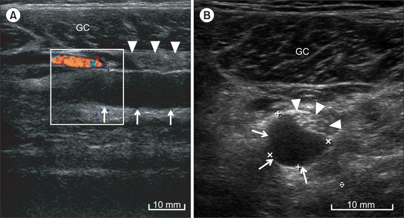
Fig. 1 (A) Longitudinal and (B) transverse ultrasonographic imaging of the lower leg showed hypoechoic cyst (arrows) along the tibial nerve (arrow heads). GC, gastrocnemius.
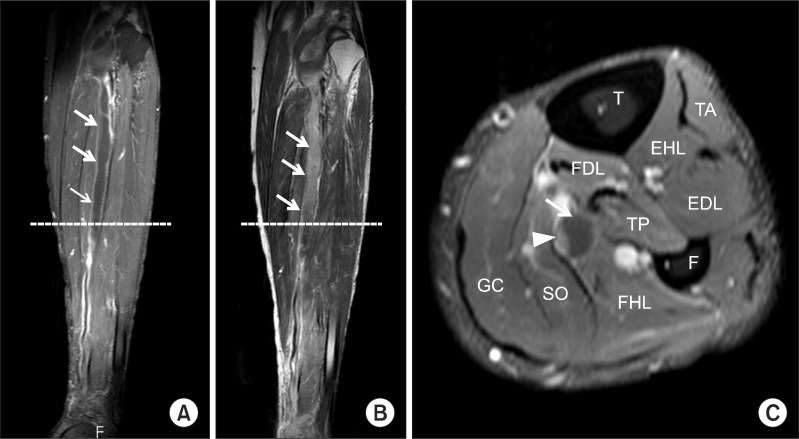
Fig. 2 (A) T1-weighted contrast enhanced coronal and (B) T2-weighted coronal magnetic resonance imaging (MRI) of the lower leg showed an elongated cystic lesion (arrows) with peripheral enhancement adjacent to the tibial nerve. (C) T1-weigted contrast enhanced transverse MRI of the lower leg showed tibial nerve (arrow head) entrapped by the cyst (arrow). T, tibia; F, fibula; TA, tibialis anterior; EHL, extensor hallucis longus; EDL, extensor digitorum longus; FDL, flexor digitorum longus; TP, tibialis posterior; FHL, flexor hallucis longus; SO, soleus; GC, gastrocnemius.
Reference
-
1. Handy JR. Popliteal cysts in adults: a review. Semin Arthritis Rheum. 2001; 31:108–118. PMID: 11590580.
Article2. Sanchez JE, Conkling N, Labropoulos N. Compression syndromes of the popliteal neurovascular bundle due to Baker cyst. J Vasc Surg. 2011; 54:1821–1829. PMID: 21958564.
Article3. Dash S, Bheemreddy SR, Tiku ML. Posterior tibial neuropathy from ruptured Baker's cyst. Semin Arthritis Rheum. 1998; 27:272–276. PMID: 9572709.
Article4. Ji JH, Shafi M, Kim WY, Park SH, Cheon JO. Compressive neuropathy of the tibial nerve and peroneal nerve by a Baker's cyst: case report. Knee. 2007; 14:249–252. PMID: 17300942.
Article5. Scott WN, Jacobs B, Lockshin MD. Posterior compartment syndrome resulting from a dissecting popliteal cyst: case report. Clin Orthop Relat Res. 1977; (122):189–192. PMID: 837605.
- Full Text Links
-
- Actions
-
Cited
- CITED
-
- Close
- Share
-
- Similar articles
-
- A Baker's Cyst Causing Common Peroneal Nerve and Tibial Nerve Entrapment Neuropathy : A Case Report
- Posterior tibial neuropathy by a Baker's cyst
- Baker's Cyst Filled with Hematoma at the Lower Calf
- Calf “Arch Sign†Seen on a Tc-99m-MDP Bone Scan Is Indicative of Synovial Fluid Leak in Ruptured Baker's Cysts: Case Reports and Analysis of Literature
- Baker's Cyst with Intramuscular Extension into Vastus Medialis Muscle
