Ann Dermatol.
2010 Nov;22(4):431-434. 10.5021/ad.2010.22.4.431.
A Case of Trichogerminoma
- Affiliations
-
- 1Department of Dermatology, Seoul National University College of Medicine, Seoul, Korea.
- 2Department of Dermatology, Seoul National University Boramae Hospital, Seoul, Korea. sycho@snu.ac.kr
- KMID: 2266181
- DOI: http://doi.org/10.5021/ad.2010.22.4.431
Abstract
- Trichogerminoma is a rare neoplasm which was first described in 1992 and there is still controversy over its inclusion into the spectrum of trichoblastoma. A 79-year-old woman presented with a 5-year history of an asymptomatic nodule on the left posterior neck. Histologically, the lesion revealed a well-demarcated deep dermal nodule surrounded by a pseudocapsule. The tumor was composed of lobules with basophilic cells and some of the lobules displayed a distinctive pattern of densely packed 'cell balls' with peripheral condensation. Immunohistochemically, the tumor cells showed zonal CK5/6 immunoactivity in contrast with the negatively stained 'cell balls'. These characteristics were compatible with the diagnosis of trichogerminoma. We report here on a rare case of a hair germ tumor called trichogerminoma.
Keyword
Figure
-
Fig. 1 A solitary non-ulcerated, hemispheric, well-demarcated, flesh-colored nodule on the posterior neck
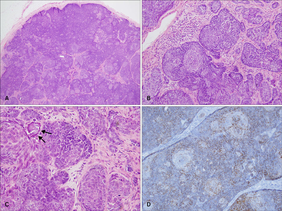
Fig. 2 (A) A well-demarcated tumor composed of basaloid lobules surrounded by a pseudocapsule in the deep dermis (H&E, ×40). (B) Distinctive 'cell balls' of pale cells with peripheral condensation (H&E, ×200). (C) Keratinous microcysts (arrow) are located in the eccentric part of the cell ball. The tumor cells extending from lobules make anastomosing strands or buds (H&E, ×400). (D)The peripheral rim of the cell balls were prominently stained with anti-CK5/6, whereas the centrally located pale 'cell balls' expressed weaker staining with anti-CK5/6 (avidin-biotin complex, ×200)
Reference
-
1. Sau P, Lupton GP, Graham JH. Trichogerminoma. Report of 14 cases. J Cutan Pathol. 1992. 19:357–365.
Article2. Kazakov DV, Kutzner H, Rutten A, Dummer R, Burg G, Kempf W. Trichogerminoma: a rare cutaneous adnexal tumor with differentiation toward the hair germ epithelium. Dermatology. 2002. 205:405–408.
Article3. Tellechea O, Reis JP. Trichogerminoma. Am J Dermatopathol. 2009. 31:480–483.
Article4. Pozo L, Diaz-Cano SJ. Trichogerminoma: further evidence to support a specific follicular neoplasm. Histopathology. 2005. 46:108–110.
Article5. Moll R, Divo M, Langbein L. The human keratins: biology and pathology. Histochem Cell Biol. 2008. 129:705–733.6. Ivan D, Hafeez Diwan A, Prieto VG. Expression of p63 in primary cutaneous adnexal neoplasms and adenocarcinoma metastatic to the skin. Mod Pathol. 2005. 18:137–142.7. Schulz T, Hartschuh W. Merkel cells are absent in basal cell carcinomas but frequently found in trichoblastomas. An immunohistochemical study. J Cutan Pathol. 1997. 24:14–24.8. Ackerman AB, de Viragh PA, Chongchitnant N. Ackerman AB, de Viragh PA, Chongchitnant N, editors. Trichoblastoma. Neoplasms with follicular differentiation. 1993. Philadelphia: Lea & Febiger;359–422.9. Kurzen H, Esposito L, Langbein L, Hartschuh W. Cytokeratins as markers of follicular differentiation: an immunohistochemical study of trichoblastoma and basal cell carcinoma. Am J Dermatopathol. 2001. 23:501–509.10. Rapini RP. Rapini RP, editor. Follicular neoplasm. Practical dermatopathology. 2005. Philadelphia: Elsevier Mosby;285–293.
