Ann Dermatol.
2013 Feb;25(1):80-83. 10.5021/ad.2013.25.1.80.
New World Cutaneous Leishmaniasis Treated with Intralesional Injection of Pentavalent Antimony
- Affiliations
-
- 1Department of Dermatology, College of Medicine, The Catholic University of Korea, Seoul, Korea. hjpark@catholic.ac.kr
- KMID: 2265975
- DOI: http://doi.org/10.5021/ad.2013.25.1.80
Abstract
- Cutaneous leishmaniasis is a skin infection caused by the Leishmania species, an intracellular protozoan parasite that is transmitted by various species of female sandflies. According to the geographic distribution and vectors, leishmaniasis is classified as Old World or New World cutaneous leishmaniasis. In Korea, 24 cases of Old World cutaneous leishmaniasis have been reported, but New World cutaneous leishmaniasis has not been reported as yet. A 37-year-old man presented with a 3-month history of a painful and erythematous nodule with two satellite papules on the left postauricular area and a papule on the left arm after traveling to the Amazon region in Brazil. After we performed skin biopsies of the lesions, diagnosis of cutaneous leishmaniasis was made by the histopathological findings. After intralesional injection of sodium stibogluconate (Pentostam(R), GlaxoSmithKline) twice a week for 4 weeks, the lesions improved with scarring. Herein, we discuss this case of New World cutaneous leishmaniasis that was successfully treated with intralesional injection of sodium stibogluconate (Pentostam(R)) in Korea.
MeSH Terms
Figure
-

Fig. 1 (A) A solitary, painful, and erythematous nodule with two satellite papules on the left postauricular area and (B) a papule on the left upper arm.
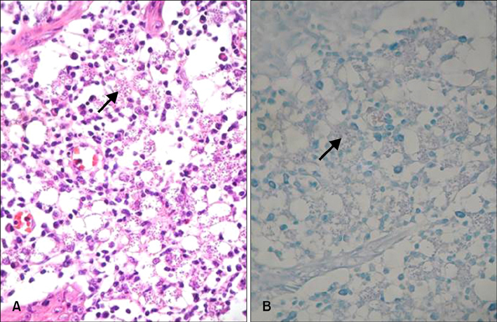
Fig. 2 Histopathological findings show numerous Leishman-Donovan bodies (arrows) in the cytoplasm of macrophages (A: H&E stain, ×400; B: Giemsa stain, ×400).

Fig. 3 Histopathological findings show chronic granulomatous inflammation and no evidence of Leishman-Donovan bodies (A: H&E stain, ×100; B: H&E stain, ×400).
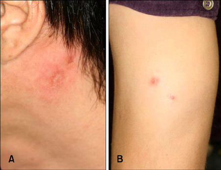
Fig. 4 Skin lesions at 3 months after treatment (A) on left postauricular area and (B) left arm.
Reference
-
1. Hepburn NC. Cutaneous leishmaniasis. Clin Exp Dermatol. 2000. 25:363–370.
Article2. Grevelink SA, Lerner EA. Leishmaniasis. J Am Acad Dermatol. 1996. 34:257–272.
Article3. Kim YJ, Hwang ES, Yoo DS, Son SW, Uhm CS, Kim IH. A case of localized cutaneous leishmaniasis in a native Korean. Korean J Dermatol. 2004. 42:884–888.4. David CV, Craft N. Cutaneous and mucocutaneous leishmaniasis. Dermatol Ther. 2009. 22:491–502.
Article5. Dowlati Y. Cutaneous leishmaniasis: clinical aspect. Clin Dermatol. 1996. 14:425–431.6. Machado P, Araújo C, Da Silva AT, Almeida RP, D'Oliveira A Jr, Bittencourt A, et al. Failure of early treatment of cutaneous leishmaniasis in preventing the development of an ulcer. Clin Infect Dis. 2002. 34:E69–E73.
Article7. Herwaldt BL. Leishmaniasis. Lancet. 1999. 354:1191–1199.
Article8. Scope A, Trau H, Anders G, Barzilai A, Confino Y, Schwartz E. Experience with New World cutaneous leishmaniasis in travelers. J Am Acad Dermatol. 2003. 49:672–678.
Article9. Seaton RA, Morrison J, Man I, Watson J, Nathwani D. Out-patient parenteral antimicrobial therapy--a viable option for the management of cutaneous leishmaniasis. QJM. 1999. 92:659–667.
Article10. Weigle KA, de Dávalos M, Heredia P, Molineros R, Saravia NG, D'Alessandro A. Diagnosis of cutaneous and mucocutaneous leishmaniasis in Colombia: a comparison of seven methods. Am J Trop Med Hyg. 1987. 36:489–496.
Article11. Marfurt J, Niederwieser I, Makia ND, Beck HP, Felger I. Diagnostic genotyping of Old and New World Leishmania species by PCR-RFLP. Diagn Microbiol Infect Dis. 2003. 46:115–124.
Article12. Weina PJ, Neafie RC, Wortmann G, Polhemus M, Aronson NE. Old world leishmaniasis: an emerging infection among deployed US military and civilian workers. Clin Infect Dis. 2004. 39:1674–1680.
Article13. Palumbo E. Current treatment for cutaneous leishmaniasis: a review. Am J Ther. 2009. 16:178–182.
Article14. Choi HM, Myung KB, Kook HI. A case of cutaneous leishmaniasis treated with cryosurgery. Korean J Dermatol. 1983. 21:207–211.15. Oliveira-Neto MP, Schubach A, Mattos M, da Costa SC, Pirmez C. Intralesional therapy of American cutaneous leishmaniasis with pentavalent antimony in Rio de Janeiro, Brazil--an area of Leishmania (V.) braziliensis transmission. Int J Dermatol. 1997. 36:463–468.
Article
- Full Text Links
-
- Actions
-
Cited
- CITED
-
- Close
- Share
-
- Similar articles
-
- A Case of Cutaneous Leishmaniasis Treated with Intralesional Injection of Meglumine Antimoniate
- A Case of Cutaneous Leishmaniasis Treated with Cryosurgery
- A Case of Scrotal Eczema with Calcified Nodules
- Cutaneous Leishmaniasis Treated with Metronidazole and Cryotherapy
- Evaluation of the Interventional Approaches in the Management of Cutaneous Leishmaniasis in Jazan: An Observational Study
