Ann Dermatol.
2013 Aug;25(3):348-351. 10.5021/ad.2013.25.3.348.
A Case of Eccrine Porocarcinoma: Usefulness of Immunostain for S-100 Protein in the Diagnoses of Recurrent and Metastatic Dedifferentiated Lesions
- Affiliations
-
- 1Department of Pathology, Osaka Medical College, Takatsuki, Japan. pa1021@art.osaka-med.ac.jp
- KMID: 2265876
- DOI: http://doi.org/10.5021/ad.2013.25.3.348
Abstract
- Eccrine porocarcinoma is a rare malignant tumor. Immunostain for S-100 protein, in addition to epithelial membrane antigen (EMA) and carcinoembryonic antigen (CEA), is described to be useful in the diagnosis. Herein, we report a case of eccrine porocarcinoma with immunostain for S-100 protein which was useful in diagnoses of recurrent and metastatic lesions. The primary lesion in the left inguinal region was excised, but it recurred on the same site 14 months after the resection. The recurrent lesion showed epithelioid melanocytic findings. Three months later, metastasis to the lungs was found. Since these recurrent and metastatic lesions were dedifferentiated, typical histologic findings of eccrine porocarcinoma disappeared in biopsied specimens. Nevertheless, scattered immunoreactive cells for S-100 protein were maintained in these dedifferentiated lesions. S-100 protein positive cells could be an aid to diagnose, even if histologic findings of recurrent and metastatic lesions have changed by dedifferentiation.
MeSH Terms
Figure
-
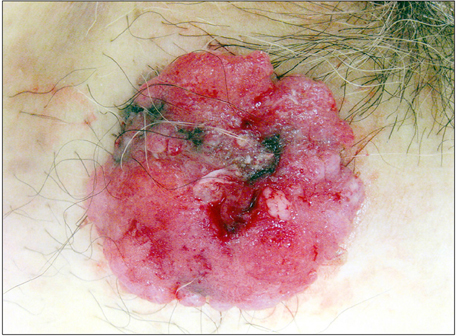
Fig. 1 Fungated polypoid lesion in the inguinal lesion.

Fig. 2 Protruded elevation is formed by the lobulated infiltrative neoplasm connected to the epidermis. Tumor cells make small glandular structures (H&E, ×100).

Fig. 3 Immunoreactivity for carcinoembryonic antigen is found at the rims of lumens (×200).
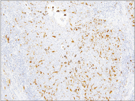
Fig. 4 Immunostain for S-100 protein reveals scattered positive cells in the primary lesion (×200).

Fig. 5 Histologic findings of recurrent lesions. Epithelioid cells proliferate diffusely in the subcutaneous tissue (H&E, ×200).

Fig. 6 Immunostain for S-100 protein reveals scattered positive cells in the recurrent lesion (×200).
Reference
-
1. Requena L, Kutzner H, Hurt MA, SantaCruz DJ, Mehregan DA, Mehregan DR, et al. Malignant tumours with apocrine and eccrine differentiation. In : Leboit PE, Burg G, Weedon D, Sarasin A, editors. Pathology and genetics of skin tumours. World Health Organization classification of tumours. New York: IARC Press;2006. p. 125–138.2. Shiohara J, Koga H, Uhara H, Takata M, Saida T. Eccrine porocarcinoma: clinical and pathological studies of 12 cases. J Dermatol. 2007; 34:516–522.
Article3. Robson A, Greene J, Ansari N, Kim B, Seed PT, McKee PH, et al. Eccrine porocarcinoma (malignant eccrine poroma): a clinicopathologic study of 69 cases. Am J Surg Pathol. 2001; 25:710–720.4. Wollina U, Castelli E, Rülke D. Immunohistochemistry of eccrine poroma and porocarcinoma--more than acrosyringeal tumors? Recent Results Cancer Res. 1995; 139:303–316.5. Nakanishi Y, Matsuno Y, Shimoda T, Wada T, Yamazaki N, Yamamoto A, et al. Eccrine porocarcinoma with melanocyte colonization. Br J Dermatol. 1998; 138:519–521.
Article6. Hara K, Kamiya S. Pigmented eccrine porocarcinoma: a mimic of malignant melanoma. Histopathology. 1995; 27:86–88.
Article7. Maeda T, Mori H, Matsuo T, Nakagawa J, Yamauchi H, Tuziguchi H, et al. Malignant eccrine poroma with multiple visceral metastases: report of a case with autopsy findings. J Cutan Pathol. 1996; 23:566–570.
Article8. Shrestha P, Muramatsu Y, Kudeken W, Mori M, Takai Y, Ilg EC, et al. Localization of Ca(2+)-binding S100 proteins in epithelial tumours of the skin. Virchows Arch. 1998; 432:53–59.
Article9. Nascimento AG. Dedifferentiated liposarcoma. Semin Diagn Pathol. 2001; 18:263–266.10. Roy P, Bullock MJ, Perez-Ordoñez B, Dardick I, Weinreb I. Epithelial-myoepithelial carcinoma with high grade transformation. Am J Surg Pathol. 2010; 34:1258–1265.
Article
- Full Text Links
-
- Actions
-
Cited
- CITED
-
- Close
- Share
-
- Similar articles
-
- Two Cases of Eccrine Porocarcinoma
- A Case of Malignant Hidracanthoma Simplex Showing Borst-Jadassohn Phenomenon
- A Case of Eccrine Porocarcinoma That Showed Squamous Differentiation on the Palm
- A Case of Eccrine Porocarcinoma, Misdiagnosed in Initial Biopsy but Diagnosed with Re-Examination by Dermoscopy
- A Case of Eccrine Porocarcinoma
