Epidermotropic Metastatic Melanoma Clinically Resembling Agminated Spitz Nevi
- Affiliations
-
- 1Department of Dermatology, Korea University Ansan Hospital, Ansan, Korea. kumcihk@korea.ac.kr
- KMID: 2265502
- DOI: http://doi.org/10.5021/ad.2014.26.5.628
Abstract
- Herein, we report a 36-year-old Asian male patient who presented with grouped multiple erythematous waxy papules and nodules on his right medial thigh. He had undergone amputation of the right second toe because of a stage IIa malignant melanoma, 3 years previously. At the time of surgery for the primary tumor, right inguinal lymph node dissection revealed no nodal involvement. Three years after the diagnosis of the primary tumor, crops of multiple erythematous papules and nodules developed. Initial histopathologic evaluation of the papules showed nests of small epithelioid cells similar to compound nevi. However, cytologic features, including high mitotic figures, lack of maturation, and some hyperchromatic nuclei suggested metastatic melanoma. In addition to the pathologic findings, the tumors were on the right thigh, which was the same side as the primary malignant melanoma. The patient underwent wide excision of the tumor and split-thickness skin grafting.
MeSH Terms
Figure
-
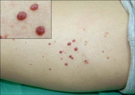
Fig. 1 Multiple erythematous waxy papules and nodules on the right medial thigh (insert: close-up view).
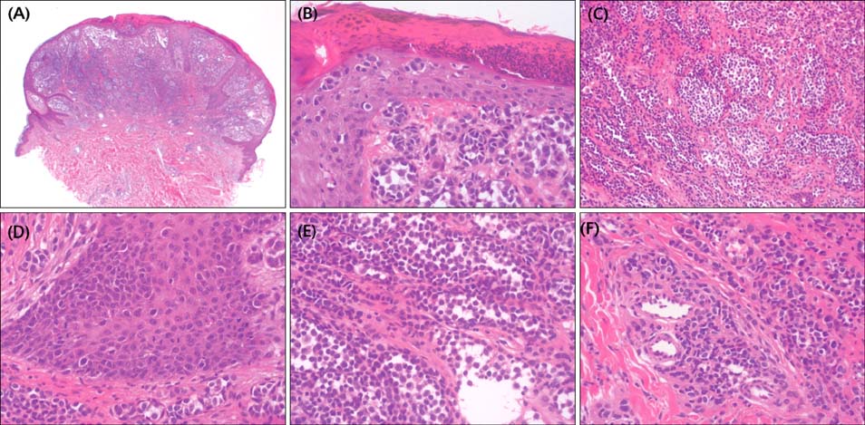
Fig. 2 (A) Scanning view showing a well-circumscribed symmetric dome-shaped nodule comprising various-sized nests of tumor cells (H&E, ×12). (B) Epidermal tumor nests and single cell exhibiting pagetoid spread throughout the epidermis (H&E, ×40). (C) Tumor cells exhibiting epithelioid morphology, with monotonous and hyperchromatic nuclei, and loss of maturation (H&E, ×100). (D) Atypical mitoses in the epidermis (H&E, ×200). (E) Atypical mitoses in the dermis (H&E, ×200). (F) Atypical tumor cell nests and individual cells around blood vessels and inside vessel lumens, indicating angiotropism (H&E, ×200).

Fig. 3 Immunohistochemical analysis. (A) Ki-67 index <5%; (B) p53, (C) HMB-45, and (D) Cyclin D1 are focally positive on the superficial dermis; (E) p16 is only sparsely positive throughout the lesion; (F) Melan-A and (G) Bcl-2 are diffusely positive in the lesion (×100). HMB-45: human melanoma black-45.
Reference
-
1. Plaza JA, Torres-Cabala C, Evans H, Diwan HA, Suster S, Prieto VG. Cutaneous metastases of malignant melanoma: a clinicopathologic study of 192 cases with emphasis on the morphologic spectrum. Am J Dermatopathol. 2010; 32:129–136.
Article2. Kato T, Demitsu T, Tomita Y, Tagami H. New primary malignant melanoma, epidermotropism and Indian-file arrangement of metastatic tumor cells in a case with intransit metastases of acral type of malignant melanoma. Dermatologica. 1986; 173:95–100.
Article3. Barnhill RL. Dermatopathology. 3rd ed. New York: McGraw-Hill Medical;2010.4. Balch CM, Buzaid AC, Soong SJ, Atkins MB, Cascinelli N, Coit DG, et al. Final version of the American Joint Committee on Cancer staging system for cutaneous melanoma. J Clin Oncol. 2001; 19:3635–3648.
Article5. Kornberg R, Harris M, Ackerman AB. Epidermotropically metastatic malignant melanoma. Differentiating malignant melanoma metastatic to the epidermis from malignant melanoma primary in the epidermis. Arch Dermatol. 1978; 114:67–69.
Article6. Bengoechea-Beeby MP, Velasco-Osés A, Mouriño Fernández F, Reguilón-Rivero MC, Remón-Garijo L, Casado-Pérez C. Epidermotropic metastatic melanoma. Are the current histologic criteria adequate to differentiate primary from metastatic melanoma? Cancer. 1993; 72:1909–1913.
Article7. Jackson R. Epidermotropic malignant melanoma: the distinction between metastatic and new primary lesions in the skin. Can J Surg. 1984; 27:533–535.8. White WL, Hitchcock MG. Dying dogma: the pathological diagnosis of epidermotropic metastatic malignant melanoma. Semin Diagn Pathol. 1998; 15:176–188.9. Gerami P, Shea C, Stone MS. Angiotropism in epidermotropic metastatic melanoma: another clue to the diagnosis. Am J Dermatopathol. 2006; 28:429–433.10. Bastian BC, Lazar A. Melanoma. In : Calonje E, McKee PH, editors. McKee's pathology of the skin. 4th ed. Edinburgh: Saunders;2011. p. 1221–1267.11. Inohara S, Kitagawa K, Kitano Y. Expression of cyclin d1 and p53 protein in various malignant skin tumors. Dermatology. 1996; 192:94–98.
Article12. McNeer G, Dasgupta T. Life history of melanoma. Am J Roentgenol Radium Ther Nucl Med. 1965; 93:686–694.13. Petersen NC, Bodenham DC, Lloyd OC. Malignant melanomas of the skin. A study of the origin, development, aetiology, spread, treatment, and rognosis. I. Br J Plast Surg. 1962; 15:49–94.14. Zbytek B, Carlson JA, Granese J, Ross J, Mihm MC Jr, Slominski A. Current concepts of metastasis in melanoma. Expert Rev Dermatol. 2008; 3:569–585.
Article
- Full Text Links
-
- Actions
-
Cited
- CITED
-
- Close
- Share
-
- Similar articles
-
- A Case of Multiple Agminated Spitz Nevi Showing Desmoplastic Changes
- A Case of Speckled Lentiginous Nevus combined with Spitz Nevi
- A Study on Immunohistochemical Stain for S-100 Protein, HMB 45 and Proliferating Cell Nuclear Antigen(PCNA) of Spitz Nevus Compared with Benign Nevus and Malignant Melanoma
- A Case of Pigmented Spitz Nevus in a Child
- Case Reports of Spitz Nevus with a Halo Phenomenon
