Ann Dermatol.
2012 Aug;24(3):306-310. 10.5021/ad.2012.24.3.306.
Clinical Experience in the Treatment of Port-Wine Stains with Blebs
- Affiliations
-
- 1Department of Dermatology, Eulji Hospital, College of Medicine, Eulji University, Seoul, Korea. drhams@eulji.ac.kr
- 2S&U Dermatology Clinic, Seoul, Korea.
- KMID: 2265303
- DOI: http://doi.org/10.5021/ad.2012.24.3.306
Abstract
- BACKGROUND
The current modality of choice for the treatment of Port-wine stains (PWS) is laser photocoagulation. Laser therapy for the treatment of PWS, especially with a pulsed dye laser (PDL), has been proven safe and effective; however, because penetration of the PDL is too shallow for an effective ablation of the blebs, treatment of blebbed PWS, using PDL, may be insufficient.
OBJECTIVE
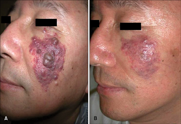
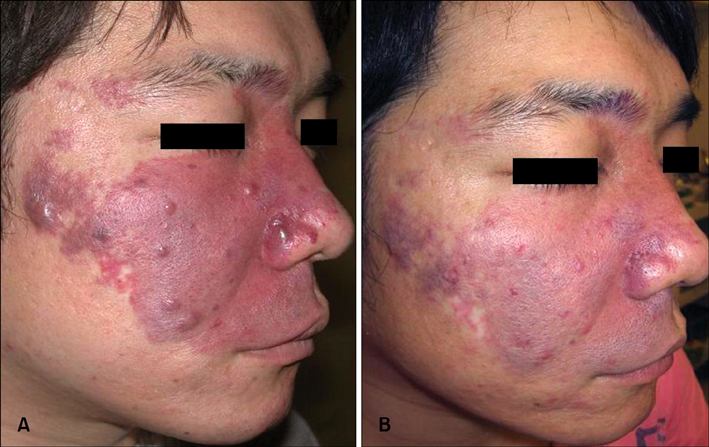
We demonstrated the clinical efficacy of a 1,064 nm long pulsed Nd:YAG laser with contact cooling device for blebbed PWS.
METHODS
Twenty one patients with blebbed PWS (Fitzpatrick skin types II-V) underwent a treatment, using a 1,064 nm long pulsed Nd:YAG laser with a contact cooling device at 8-week intervals. Treatments were done using 5~6 mm spot sizes at 20~30 ms and 95~170 J/cm2. Laser parameters were adjusted in order to meet the needs of each individual patient's lesions.
RESULTS
All subjects tolerated the treatments well, and showed clinical improvement from blebs. Of the 21 patients, 18 of them experienced either moderate or excellent response.
CONCLUSION
Use of a 1,064 nm long pulsed Nd:YAG laser results in a greater depth of vascular coagulation. A 1,064 nm long pulsed Nd:YAG laser with contact cooling device may be regarded as a promising therapeutic option for the treatment of blebbed PWS.
Figure
Reference
-
1. Adamic M, Troilius A, Adatto M, Drosner M, Dahmane R. Vascular lasers and IPLS: guidelines for care from the European Society for Laser Dermatology (ESLD). J Cosmet Laser Ther. 2007. 9:113–124.
Article2. Lanigan SW, Taibjee SM. Recent advances in laser treatment of port-wine stains. Br J Dermatol. 2004. 151:527–533.
Article3. Chapas AM, Geronemus RG. Physiologic changes in vascular birthmarks during early infancy: mechanisms and clinical implications. J Am Acad Dermatol. 2009. 61:1081–1082.
Article4. Ahcan U, Zorman P, Recek D, Ralca S, Majaron B. Port wine stain treatment with a dual-wavelength Nd:Yag laser and cryogen spray cooling: a pilot study. Lasers Surg Med. 2004. 34:164–167.
Article5. Stier MF, Glick SA, Hirsch RJ. Laser treatment of pediatric vascular lesions: Port wine stains and hemangiomas. J Am Acad Dermatol. 2008. 58:261–285.
Article6. Groot D, Rao J, Johnston P, Nakatsui T. Algorithm for using a long-pulsed Nd:YAG laser in the treatment of deep cutaneous vascular lesions. Dermatol Surg. 2003. 29:35–42.
Article7. Dover JS, Sadick NS, Goldman MP. The role of lasers and light sources in the treatment of leg veins. Dermatol Surg. 1999. 25:328–335.
Article8. Gilchrest BA, Rosen S, Noe JM. Chilling port wine stains improves the response to argon laser therapy. Plast Reconstr Surg. 1982. 69:278–283.
Article9. Edström DW, Hedblad MA, Ros AM. Flashlamp pulsed dye laser and argon-pumped dye laser in the treatment of port-wine stains: a clinical and histological comparison. Br J Dermatol. 2002. 146:285–289.
Article10. Frohm Nilsson M, Passian S, Wiegleb Edstrom D. Comparison of two dye lasers in the treatment of port-wine stains. Clin Exp Dermatol. 2010. 35:126–130.
Article11. Yang MU, Yaroslavsky AN, Farinelli WA, Flotte TJ, Rius-Diaz F, Tsao SS, et al. Long-pulsed neodymium:yttrium-aluminum-garnet laser treatment for port-wine stains. J Am Acad Dermatol. 2005. 52(3 Pt 1):480–490.
Article12. Haedersdal M, Efsen J, Gniadecka M, Fogh H, Keiding J, Wulf HC. Changes in skin redness, pigmentation, echostructure, thickness, and surface contour after 1 pulsed dye laser treatment of port-wine stains in children. Arch Dermatol. 1998. 134:175–181.
Article13. Nguyen CM, Yohn JJ, Huff C, Weston WL, Morelli JG. Facial port wine stains in childhood: prediction of the rate of improvement as a function of the age of the patient, size and location of the port wine stain and the number of treatments with the pulsed dye (585 nm) laser. Br J Dermatol. 1998. 138:821–825.
Article14. Namba Y, Mae O, Ao M. The treatment of port wine stains with a dye laser: a study of 644 patients. Scand J Plast Reconstr Surg Hand Surg. 2001. 35:197–202.
Article
- Full Text Links
- Actions
-
Cited
- CITED
-
- Close
- Share
- Similar articles
-
- The combined application of bleomycin with triamcinolone acetonide in port wine stains: inhibiting proliferation and recurrence of port wine stains
- Five Cases of Acquired Port-Wine Stains
- A Case of Granuloma Pyogenicum within a Port-wine Stain
- Treatment of Port Wine Stain with Nodular Change
- Treatment Using a Long Pulsed Nd:Yag Laser with a Pulsed Dye Laser for Four Cases of Blebbed Port Wine Stains


