Ann Dermatol.
2015 Apr;27(2):237-238. 10.5021/ad.2015.27.2.237.
A Case of Keratoacanthoma Associated with Basal Cell Carcinoma
- Affiliations
-
- 1Department of Dermatology, Gangnam Severance Hospital, Cutaneous Biology Research Institute, Yonsei University College of Medicine, Seoul, Korea. karenroh@yuhs.ac
- KMID: 2264827
- DOI: http://doi.org/10.5021/ad.2015.27.2.237
Abstract
- No abstract available.
MeSH Terms
Figure
-
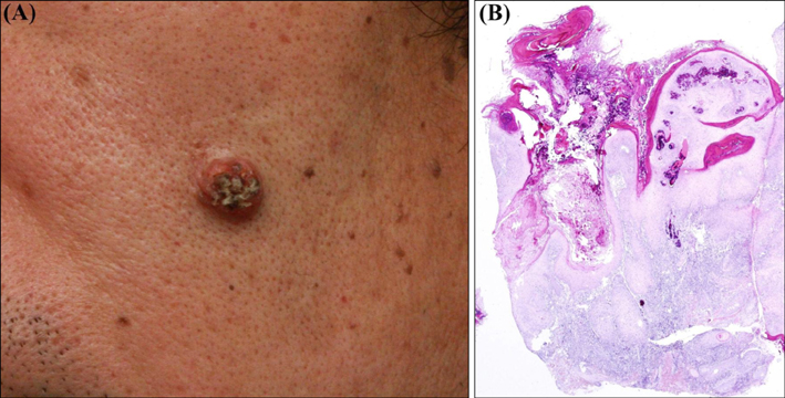
Fig. 1 (A) A 6-mm-sized, protruding, verrucous papule on the left cheek. (B) Central keratotic plug surrounded by epithelial proliferation (H&E, ×20).
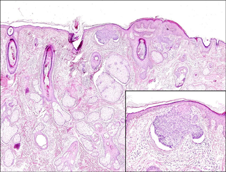
Fig. 2 Single tumor nest with abundant sebaceous glands and hair follicles (H&E, ×100). Basal cell carcinoma showing palisading basaloid cells with a cleft between the tumor nest and stroma (inset, ×200).
Reference
-
1. Einaugler RB, Henkind P, De Oliveira LF, Bart RS. Keratoacanthoma with basal cell carcinoma. Am J Ophthalmol. 1968; 65:922–925.
Article2. Butcher RB 2nd. Malignant potential of keratoacanthoma. Laryngoscope. 1979; 89:1092–1098.
Article3. Bryant J. Basal cell carcinoma associated with keratoacanthoma. J Dermatol Surg Oncol. 1985; 11:1230–1231.
Article4. Ahlgrimm-Siess V, Hofmann-Wellenhof R, Zalaudek I, Cerroni L, Kerl H. Collision of malignant melanoma (lentigo maligna type) with squamous cell carcinoma in solar-damaged skin of the face. Dermatol Surg. 2007; 33:122–124.
Article5. Kim J, Roh HJ, Chung KY, Roh MR. Collision of two rare adnexal tumors with folliculosebaceous differentiation. J Am Acad Dermatol. 2011; 64:e84–e85.
Article
