A Case of Spitzoid Melanoma
- Affiliations
-
- 1Department of Dermatology, Hallym University Sacred Heart Hospital, Hallym University College of Medicine, Anyang, Korea. dermakkh@naver.com
- KMID: 2264813
- DOI: http://doi.org/10.5021/ad.2015.27.2.206
Abstract
- Spitzoid melanoma is a subtype of melanoma that, clinically and histologically, resembles a Spitz nevus. Clinically, spitzoid melanomas usually evolve from amelanotic nodular lesions, growing to 1 cm or more in diameter. They often remain clinically undiagnosed because of their wide variety of clinical appearances and a lack of pigmentation. Distinguishing a Spitz nevus from a spitzoid melanoma can be extremely difficult. Features that favor the diagnosis of a spitzoid melanoma are asymmetrical shape, diameter greater than 1 cm, a lesion with a deep invasive component, and a high degree of cytologic atypia. There have been only rare reports in the literature of the presence of giant cells in malignant melanoma, and the presence of these cells may result in its misdiagnosis as a histiocytic tumor. We present a case of spitzoid melanoma on the right ankle of a 22-year-old-woman.
Keyword
MeSH Terms
Figure
-
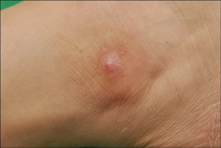
Fig. 1 Solitary, relatively ill-defined, 1.5×1 cm-sized erythematous nodule on the right ankle.
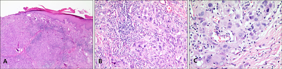
Fig. 2 (A) Epithelial granuloma-like tumor in the dermis. Atypical mitosis was found predominantly on the deep dermis, but also a little on the superficial dermis. Kamino bodies and pagetoid spread were absent from the epidermal component. The deep dermal invasion pattern of the lower border showed the mixed type, both expansive and infiltrative types (H&E, ×40). (B) The tumor cells were multinucleate and very pleomorphic with abundant cytoplasm and large vesicular nuclei (H&E, ×200). (C) Large vesicular nuclei containing prominent eosinophilic nucleoli (H&E, ×400).
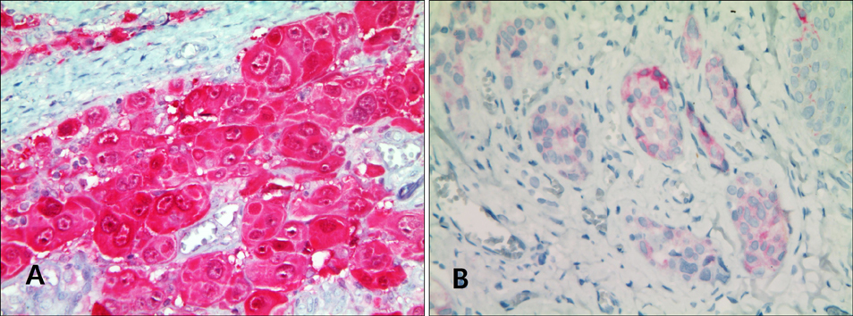
Fig. 3 (A) Positive staining reaction for S-100 (×400). (B) Positive staining reaction for Melan-A (×400).
Reference
-
1. Kamino H. Spitzoid melanoma. Clin Dermatol. 2009; 27:545–555.
Article2. Barnhill RL, Gupta K. Unusual variants of malignant melanoma. Clin Dermatol. 2009; 27:564–587.
Article3. Requena C, Botella R, Nagore E, Sanmartín O, Llombart B, Serra-Guillén C, et al. Characteristics of spitzoid melanoma and clues for differential diagnosis with spitz nevus. Am J Dermatopathol. 2012; 34:478–486.
Article4. Hornick JL. Practical soft tissue pathology: a diagnostic approach. 1st ed. Philadelphia: Elsevier;2013. 279–296.5. Calonje JE, Brenn T, Lazar AJ, McKee PH. McKee's pathology of the skin. 4th ed. Philadelphia: Elsevier;2012. p. 1221–1267.6. From L, Hanna W, Kahn HJ, Gruss J, Marks A, Baumal R. Origin of the desmoplasia in desmoplastic malignant melanoma. Hum Pathol. 1983; 14:1072–1080.
Article7. Cohen LM. The starburst giant cell is useful for distinguishing lentigo maligna from photodamaged skin. J Am Acad Dermatol. 1996; 35:962–968.
Article8. Boyd AS, Wu H, Shyr Y. Monster cells in malignant melanoma. Am J Dermatopathol. 2005; 27:208–210.
Article
- Full Text Links
-
- Actions
-
Cited
- CITED
-
- Close
- Share
-
- Similar articles
-
- A Pediatric Case of Spitzoid Melanoma with Subsequent Large Lymph Node Metastasis
- A Case of Spitzoid Melanoma with Lymph Node Metastasis in a Child
- Adult Onset Atypical Spitz Nevus
- A Case Report of Spitzoid Melanoma in a Patient with Breast Cancer
- A Case of Nodular Melanoma with a Family History of Difficult Clinical Diagnosis
