Bilateral single cord of the brachial plexus in an adult female cadaver of South Indian origin
- Affiliations
-
- 1Department of Anatomy, St. John's Medical College, Bangalore, India. nachiket76@gmail.com
- KMID: 2263151
- DOI: http://doi.org/10.5115/acb.2013.46.3.223
Abstract
- The occurrence of a brachial plexus united into a single cord is very rare. During routine dissection of an elderly female cadaver, the brachial plexus united into a single cord was observed bilaterally. On the left side, C4, C5, and C6 roots combined to form the upper trunk, the C7 root continued as the middle trunk, and C8 and T1 united to form the lower trunk. All three trunks almost immediately fused to form a single cord. On the right side, C5 and C6 roots joined to form the upper trunk, which divided into anterior and posterior divisions. C7, C8, and T1 roots combined to form the lower trunk. The anterior and posterior divisions united with the lower trunk to form a single cord. On both sides, the subclavian artery was superior to the single cord. Supraclavicular brachial plexus injuries in such individuals may have serious clinical manifestations.
Keyword
Figure
-
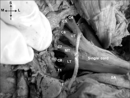
Fig. 1 Formation of the single cord on the left side. C4, C5, C6, C7, C8, T1, roots of the brachial plexus; I, inferior; L, lateral; LT, lower trunk; M, medial; MT, middle trunk; PN, phrenic nerve; S, superior; SA, subclavian artery; UT, upper trunk.

Fig. 2 The branches of the brachial plexus on the left side. AN, axillary nerve; CNA, medial cutaneous nerve of the arm; CNF, medial cutaneous nerve of the forearm; DM, deltoid muscle; I, inferior; L, lateral; M, medial; MN, median nerve; MPN, medial pectoral nerve; RN, radial nerve; S, superior; SC, single cord; ScA, displaced scalenus anterior; SN, suprascapular nerve; UN, ulnar nerve.

Fig. 3 Schematic representation of the major variations of the brachial plexus observed on the left side. B, major branches of the single cord; C4, C5, C6, C7, C8, T1, roots of the brachial plexus; LT, lower trunk; MN, median nerve; MT, middle trunk; R, roots; RN, radial nerve; SC, single cord; T, trunks; UN, ulnar nerve; UT, upper trunk.

Fig. 4 Formation of the single cord on the right side. AD, anterior division of upper trunk; AN, axillary nerve; C5, C6, C7, C8, T1, roots of the brachial plexus; I, inferior; L, lateral; M, medial; MN, median nerve; PD, posterior division of upper trunk; RN, radial nerve; S, superior; SA, subclavian artery; SC, single cord; UN, ulnar nerve.
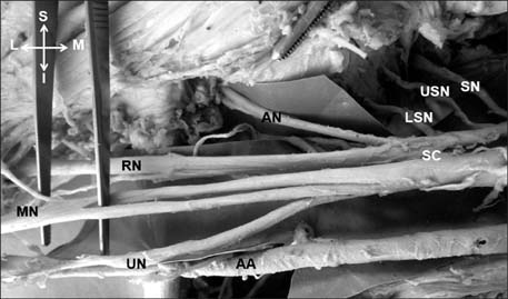
Fig. 5 Branches of the brachial plexus on the right side. AA, subclavian artery; AN, axillary nerve; I, inferior; L, lateral; LSN, lower subscapular nerve; M, medial; MN, median nerve which splits and rejoins; RN, radial nerve; S, superior; SC, single cord; SN, suprascapular nerve; UN, ulnar nerve; USN, upper subscapular nerve.
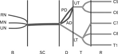
Fig. 6 Schematic representation of the major variations of the brachial plexus observed on the right side. AD, anterior division; B, major branches of the single cord; C5, C6, C7, C8, T1, roots of the brachial plexus; D, divisions; LT, lower trunk; MN, median nerve; PD, posterior division; R, roots; RN, radial nerve; SC, single cord; T, trunks; UN, ulnar nerve; UT, upper trunk.
Reference
-
1. Standring S. Gray's anatomy: the anatomical basis of clinical practice. 39th ed. Edinburgh: Elsevier Churchill Livingstone Publishers;2006.2. Khullar M, Sharma S, Khullar S. Multiple bilateral neuroanatomical variations of the nerves of the arm: a case report. Int J Med Health Sci. 2012; 1:75–84.3. Shankar N, Veeramani R. Replacement of the medial and lateral cords of the brachial plexus by a common cord and its trifurcation into major branches. Int J Anat Var. 2010; 3:205–207.4. Sweekritha , Sultana Q, Pillai V, Rao CP. A rare case of variation of the cords of brachial plexus. Int J A J Inst Med Sci. 2012; 1:75–77.5. Aggarwal A, Sahni D, Kaur H, Batra YK, Sondekoppam RV. A rare anatomical variation of the brachial plexus: single cord anomaly. Anesth Analg. 2012; 114:466–470.6. Kerr AT. The brachial plexus of nerves in man, the variations in its formation and branches. Am J Anat. 1918; 23:285–395.7. Pandey SK, Shukla VK. Anatomical variations of the cords of brachial plexus and the median nerve. Clin Anat. 2007; 20:150–156.8. Meenakshisundaram J. A single common cord in the infraclavicular part of the brachial plexus. Int J Health Sci Res. 2012; 2:108–111.9. Singer E. Human brachial plexus united into a single cord: description and interpretation. Anat Rec. 1933; 55:411–419.10. Hasan M, Narayan D. A single cord human brachial plexus. J Anat Soc India. 1964; 13:103–104.11. Iwata H. Studies on the development of the brachial plexus in Japanese embryo. Rep Dep Anat Mie Prefect Univ Sch Med. 1960; 13:129–144.12. Sannes DH, Reh TA, Harris WA. Development of the nervous system. New York: Academic Press;2000. p. 189–197.13. Williams PL, Bannister LH, Berry MM, Collins P, Dyson M, Dussek JE, Ferguson MW. Gray's anatomy. 38th ed. London: Churchill Livingstone;1999. p. 231–232.14. Miller RA. Comparative studies upon the morphology and distribution of the brachial plexus. Am J Anat. 1934; 54:143–175.15. Singhal S, Rao VV, Ravindranath R. Variations in brachial plexus and the relationship of median nerve with the axillary artery: a case report. J Brachial Plex Peripher Nerve Inj. 2007; 2:21.16. Nayak S, Somayaji N, Vollala VR, Raghunathan D, Rodrigues V, Samuel VP, Alathady Malloor P. A rare variation in the formation of the upper trunk of the brachial plexus: a case report. Neuroanatomy. 2005; 4:37–38.
- Full Text Links
-
- Actions
-
Cited
- CITED
-
- Close
- Share
-
- Similar articles
-
- Bilateral absence of musculocutaneous nerve with unusual branching pattern of lateral cord and median nerve of brachial plexus
- Bilateral Interscalene Brachial Plexus Block for Surgery on Both Upper Extremities in a Patient with Unilateral Vocal Cord Paralysis : A case report
- A Surgical Resection of Giant Schwannoma in the Brachial lexus
- Multiple unilateral variations in medial and lateral cords of brachial plexus and their branches
- Bilateral Brachial Plexopathy Following an Attempted Hanging: A Case Report
