Korean J Neurotrauma.
2014 Oct;10(2):126-129. 10.13004/kjnt.2014.10.2.126.
Visualization of a Traumatic Pseudoaneurysm at Internal Carotid Artery Bifurcation due to Blunt Head Injury: A Case Report
- Affiliations
-
- 1Department of Neurosurgery, Chonbuk National University Medical School-Hospital, Jeonju, Korea. kohejns@jbnu.ac.kr
- KMID: 2256246
- DOI: http://doi.org/10.13004/kjnt.2014.10.2.126
Abstract
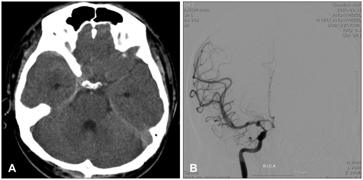
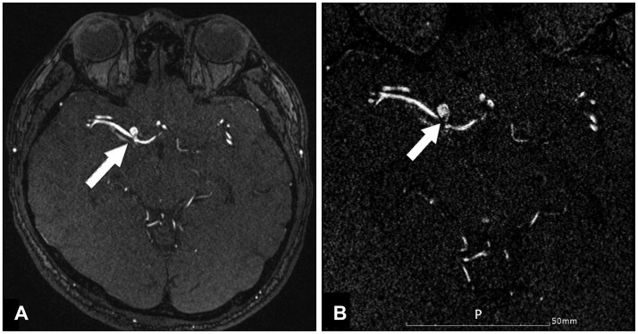
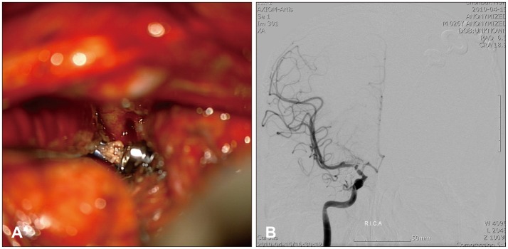
- Traumatic intracranial pseudoaneurysms occurring after blunt head injuries are rare. We report an unusual case of subarachnoid hemorrhage (SAH) caused by rupturing of the traumatic pseudoaneurysm of the internal carotid artery (ICA) bifurcation that resulted from a non-penetrating injury. In a patient with severe headache and SAH in the right sylvian cistern, which developed within 7 days after a blunt-force head injury, a trans-femoral cerebral angiogram (TFCA) showed aneurysmal sac which was insufficient to confirm the pseudoaneurysm. We obtained a multi-slab image of three dimensional time of flight (TOF) of magnetic resonance angiography (MRA). The source image of the gadolinium-enhanced MRA revealed an intimal flap within the intracranial ICA bifurcation, providing a clue for the diagnosis of a dissecting pseudoaneurysm at the ICA bifurcation due to blunt head trauma. We performed direct aneurysmal neck clipping, without neurological deficit. A follow-up TFCA did not show either aneurysm sac or luminal narrowing. We suggest that in the patient with a history of blunt head injury with SAH following shortly, multi-slab image of 3D TOF MRA can give visualization of the presence of a pseudoaneurysm.
Keyword
MeSH Terms
Figure
Reference
-
1. Acosta C, Williams PE Jr, Clark K. Traumatic aneurysms of the cerebral vessels. J Neurosurg. 1972; 36:531–536. PMID: 5026539.
Article2. Crowell RM, Oglivy CS. Traumatic intracranial aneurysms. In : Ojemann RG, Ogilvy CS, Crowell RM, Heros RC, editors. Surgical management of neuro vascular disease. Baltimore, MD: Williams & Wilkins;1995. p. 377–384.3. Dubey A, Sung WS, Chen YY, Amato D, Mujic A, Waites P, et al. Traumatic intracranial aneurysm: a brief review. J Clin Neurosci. 2008; 15:609–612. PMID: 18395452.
Article4. Fleischer AS, Patton JM, Tindall GT. Cerebral aneurysms of traumatic origin. Surg Neurol. 1975; 4:233–239. PMID: 1162596.5. Holmes B, Harbaugh RE. Traumatic intracranial aneurysms: a contemporary review. J Trauma. 1993; 35:855–860. PMID: 8263982.6. Hopkins JK, Shaibani A, Ali S, Khawar S, Parkinson R, Futterer S, et al. Coil embolization of posttraumatic pseudoaneurysm of the ophthalmic artery causing subarachnoid hemorrhage. Case report. J Neurosug. 2007; 107:1043–1046.7. Kikkawa Y, Natori Y, Sasaki T. Delayed post-traumatic pseudoaneurysmal formation of the intracranial ophthalmic artery after closed head injury. Case report. Neurol Med Chir (Tokyo). 2012; 52:41–43. PMID: 22278026.8. Kneyber MC, Rinkel GJ, Ramos LM, Tulleken CA, Braun KP. Early posttraumatic subarachnoid hemorrhage due to dissecting aneurysms in three children. Neurology. 2005; 65:1663–1665. PMID: 16301503.
Article9. Komiyama M, Morikawa T, Nakajima H, Yasui T, Kan M. "Early" apoplexy due to traumatic intracranial aneurysm--case report. Neurol Med Chir (Tokyo). 2001; 41:264–270. PMID: 11396307.10. Larson PS, Reisner A, Morassutti DJ, Abdulhadi B, Harpring JE. Traumatic intracranial aneurysms. Neurosurg Focus. 2000; 8:e4. PMID: 16906700.
Article11. Parkinson D, West M. Traumatic intracranial aneurysms. J Neurosurg. 1980; 52:11–20. PMID: 7350269.
Article12. Pozzati E, Gaist G, Servadei F. Traumatic aneurysms of the supraclinoid internal carotid artery. J Neurosurg. 1982; 57:418–422. PMID: 7097342.
Article13. Voelker JL, Ortiz O. Delayed deterioration after head trauma due to traumatic aneurysm. W V Med J. 1997; 93:317–319. PMID: 9439194.
- Full Text Links
- Actions
-
Cited
- CITED
-
- Close
- Share
- Similar articles
-
- Traumatic Pseudoaneurysm of the External and Internal Carotid Artery Presenting as Epistaxis: Case Report
- Spontaneous Recanalization from Traumatic Internal Carotid Artery Occlusion
- Internal Carotid Artery Dissection with Traumatic Pseudoaneurysm Formation after Penetrating Head Injury
- Internal Carotid Artery Pseudoaneurysm in a Patient Presenting With Recurrent Epistaxis: A Case Report and Literature Review
- Traumatic Blindness Due to Injury of Internal Carotid Artery Associated with Craniomaxillofacial Fracture


