MRI Findings of Large Low-Grade Angiosarcoma of the Breast with Subsequent Bone Metastases: A Case Report
- Affiliations
-
- 1Department of Radiology, Catholic University of the Sacred Heart-Foundation for Research and Treatment "John Paul II", Campobasso, Italy. eleonora.cucci@rm.unicatt.it
- 2Department of Oncology, Catholic University of the Sacred Heart-Foundation for Research and Treatment "John Paul II", Campobasso, Italy.
- 3Department of Radiology, Hospital "A. Cardarelli", Campobasso, Italy.
- KMID: 2242203
- DOI: http://doi.org/10.4048/jbc.2012.15.2.255
Abstract
- We describe the magnetic resonance imaging (MRI) findings of 13 cm-sized low-grade angiosarcoma of the breast that occurred in a 23-year-old woman. Magnetic resonance examination revealed an ill-defined mass with marked high-signal intensity on T2-weighted images and persistent heterogeneous enhancement. Thirty months later she developed bone metastases, incidentally found on an MRI performed to evaluate the pelvis. There were well-defined bone lesions with high-signal intensity on T2-weighted images and persistent contrast enhancement on delayed phases. The metastases were not detected on previous computed tomography and fluoro-deoxyglucose positron emission tomography scans because the lesions were subtle osteoblastic type with a low proliferative index.
Keyword
MeSH Terms
Figure
-
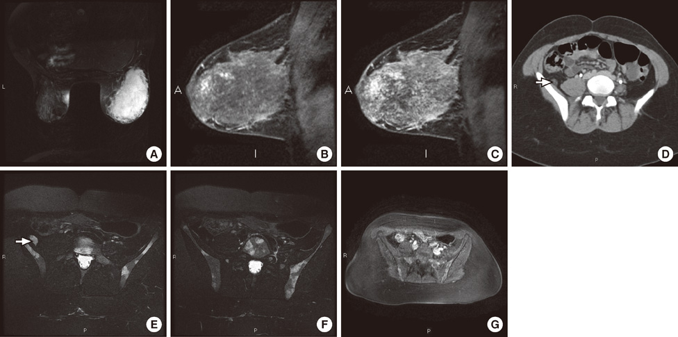
Figure 1 A low-grade angiosarcoma in a 23-year-old woman with increasing size and discomfort of the right breast. (A) Axial T2-weighted fast spin-echo magnetic resonance image shows a marked high-signal intensity mass of the right breast, mainly in the outer quadrants. (B, C) Dynamic sagittal 3D Vibrant early- (B) and late- (C) phase images show persistent heterogeneous enhancement of the mass. (D) Post-contrast computed tomography image shows suspicious right retroperitoneal pelvic lymph node (arrow). (E, F) Axial T2-weighted fast spin-echo fat saturation images confirmed retroperitoneal lymph node (E, arrow) and revealed well-defined high-signal intensity bone lesions (F). (G) Axial 3D-Liver Acquisition with Volume Acceleration images obtained late after intravenous contrast administration. The bone lesions show persistent contrast enhancement on delayed phases, suggestive of metastases.
Reference
-
1. Kikawa Y, Konishi Y, Nakamoto Y, Harada T, Takeo M, Ogata M, et al. Angiosarcoma of the breast: specific findings of MRI. Breast Cancer. 2006. 13:369–373.
Article2. Lim RF, Goei R. Best cases from the AFIP: angiosarcoma of the breast. Radiographics. 2007. 27:Suppl 1. S125–S130.3. Monroe AT, Feigenberg SJ, Mendenhall NP. Angiosarcoma after breast-conserving therapy. Cancer. 2003. 97:1832–1840.
Article4. Vorburger SA, Xing Y, Hunt KK, Lakin GE, Benjamin RS, Feig BW, et al. Angiosarcoma of the breast. Cancer. 2005. 104:2682–2688.
Article5. Yang WT, Hennessy BT, Dryden MJ, Valero V, Hunt KK, Krishnamurthy S. Mammary angiosarcomas: imaging findings in 24 patients. Radiology. 2007. 242:725–734.
Article6. Glazebrook KN, Magut MJ, Reynolds C. Angiosarcoma of the breast. AJR Am J Roentgenol. 2008. 190:533–538.
Article7. Marchant LK, Orel SG, Perez-Jaffe LA, Reynolds C, Schnall MD. Bilateral angiosarcoma of the breast on MR imaging. AJR Am J Roentgenol. 1997. 169:1009–1010.
Article8. Murakam S, Nagano H, Okubo K, Sakata H, Tsuji Y, Ishiguro T, et al. Angiosarcoma of the breast: report of a case and its findings of MRI. Breast Cancer. 2001. 8:254–258.
Article9. Sher T, Hennessy BT, Valero V, Broglio K, Woodward WA, Trent J, et al. Primary angiosarcomas of the breast. Cancer. 2007. 110:173–178.
Article10. Nakai T, Okuyama C, Kubota T, Yamada K, Ushijima Y, Taniike K, et al. Pitfalls of FDG-PET for the diagnosis of osteoblastic bone metastases in patients with breast cancer. Eur J Nucl Med Mol Imaging. 2005. 32:1253–1258.
Article11. Gatcombe HG, Olson TA, Esiashvili N. Metastatic primary angiosarcoma of the breast in a pediatric patient with a complete response to systemic chemotherapy and definitive radiation therapy: case report and review of the literature. J Pediatr Hematol Oncol. 2010. 32:192–194.
Article
