J Breast Cancer.
2012 Jun;15(2):224-229. 10.4048/jbc.2012.15.2.224.
Long-Term Follow-Up Result of Benign Phyllodes Tumor of the Breast Diagnosed and Excised by Ultrasound-Guided Vacuum-Assisted Breast Biopsy
- Affiliations
-
- 1Department of Surgery, Kangnam CHA Hospital, CHA University College of Medicine, Seoul, Korea. phl1@cha.ac.kr
- 2Department of Diagnostic Radiology, Kangnam CHA Hospital, CHA University College of Medicine, Seoul, Korea.
- 3Department of Diagnostic Pathology, Kangnam CHA Hospital, CHA University College of Medicine, Seoul, Korea.
- KMID: 2242194
- DOI: http://doi.org/10.4048/jbc.2012.15.2.224
Abstract
- PURPOSE
Percutaneous removal of benign breast tumors using ultrasound-guided vacuum-assisted breast biopsy (VABB) has been recently regarded as a feasible and safe method without serious complications. The aim of this study was to evaluate the efficacy and safety of the VABB in the treatment of benign phyllodes tumors, and to identify whether or not surgical re-excision is necessary for benign phyllodes tumors diagnosed and excised by VABB.
METHODS
From January 2003 to December 2011, a total of 6,923 VABB were performed in 5,434 patients. Out of 6,923 lesions, 53 were benign phyllodes tumors. Among these, 31 lesions, with a follow-up period of longer than 24 months, were enrolled in this study. Ultrasonography follow-up was performed at 3 to 6 month intervals in order to assess recurrence. The mean follow-up period was 75.9+/-13.5 months (range, 24-94 months).
RESULTS
The mean patient age at presentation was 31.6+/-9.4 years. The mean size of the lesion was 1.60+/-0.88 cm. The majority of lesions, 74.2% (23 cases), were palpable, and 25.8% (8 cases) were non-palpable. Twenty-two lesions (71.0%) were classified as Breast Imaging Reporting and Data System category 3, and nine lesions (29.0%) were classified as category 4a, by ultrasonography. During the follow-up period, local recurrence developed in one lesion, making the local recurrence rate 3.2%.
CONCLUSION
If a benign phyllodes tumor is diagnosed, and sufficiently excised by VABB, observing the clinical course may be considered as an alternative to performing immediate wide local excision; this is the case despite the fact that it would need to be observed for a prolonged period of time.
MeSH Terms
Figure
-

Figure 1 (A) A 1.3-cm palpable mass was located in lower outer quadrant of right breast. On ultrasonography, the lesion was a well-defined oval benign tumor of Breast Imaging Reporting and Data System (BI-RADS) category 3. The specimen was a benign biphasic tumor with a prominent stroma. The epithelial component showed adenosis. The stroma was widened and the stromal cellularity was increased. The mitoses were rare (H&E stain, B: ×40, C: ×200).
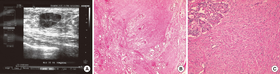
Figure 2 (A) A palpable local recurrence of Breast Imaging Reporting and Data System (BI-RADS) category 4a was found on follow-up ultrasonography. The recurred mass was also a benign biphasic tumor very similar to the primary lesion (H&E stain, B: ×40, C: ×200).
Reference
-
1. Feuer EJ, Wun LM, Boring CC, Flanders WD, Timmel MJ, Tong T. The lifetime risk of developing breast cancer. J Natl Cancer Inst. 1993. 85:892–897.
Article2. Buchanan EB. Cystosarcoma phyllodes and its surgical management. Am Surg. 1995. 61:350–355.3. Müller J. Uber den feineren Ban und Die Formen der Krankaften Geschwulste. 1838. Berlin: G. Reimer;54–57. Cited from Chen WH, Cheng SP, Tzen CY, Yang TL, Jeng KS, Liu CL, et al. Surgical treatment of phyllodes tumors of the breast: retrospective review of 172 cases. J Surg Oncol 2005;91:185-94.4. World Health Organization. Histological typing of breast tumors. Tumori. 1982. 68:181–198.5. Joshi SC, Sharma DN, Bahadur AK, Maurya R, Kumar S, Khurana N. Cystosarcoma phyllodes: our institutional experience. Australas Radiol. 2003. 47:434–437.
Article6. Deeming G, Divakaran R, Butterworth D, Foster M. Temporomandibular region metastasis from cystosarcoma phyllodes: a case report and review of the literature. J Craniomaxillofac Surg. 2003. 31:325–328.
Article7. Guerrero MA, Ballard BR, Grau AM. Malignant phyllodes tumor of the breast: review of the literature and case report of stromal overgrowth. Surg Oncol. 2003. 12:27–37.
Article8. Shabahang M, Franceschi D, Sundaram M, Castillo MH, Moffat FL, Frank DS, et al. Surgical management of primary breast sarcoma. Am Surg. 2002. 68:673–677.9. Park HL, Kwak JY, Lee SH, Kim JY, Kim KI, Kim WW, et al. Excision of benign breast disease by ultrasound-guided vacuum assisted biopsy device (Mammotome). J Korean Surg Soc. 2005. 68:96–101.10. Park HL, Kwak JY, Jung H, Lee SH, Shim JY, Kim JY, et al. Is mammotome excision feasible for benign breast mass bigger than 3 cm in greatest dimension? J Korean Surg Soc. 2006. 70:25–29.11. American College of Radiology, BI-RADS Committee. ACR BI-RADS Breast Imaging and Reporting Data System: Breast Imaging Atlas. 2003. 4th ed. Reston: American College of Radiology.12. Treves N, Sunderland DA. Cystosarcoma phyllodes of the breast: a malignant and a benign tumor: a clinicopathological study of seventy-seven cases. Cancer. 1951. 4:1286–1332.
Article13. Noguchi S, Motomura K, Inaji H, Imaoka S, Koyama H. Clonal analysis of fibroadenoma and phyllodes tumor of the breast. Cancer Res. 1993. 53:4071–4074.14. Chua CL, Thomas A, Ng BK. Cystosarcoma phyllodes: a review of surgical options. Surgery. 1989. 105(2 Pt 1):141–147.15. Ciatto S, Bonardi R, Cataliotti L, Cardona G. Phyllodes tumor of the breast: a multicenter series of 59 cases. Coordinating Center and Writing Committee of FONCAM (National Task Force for Breast Cancer), Italy. Eur J Surg Oncol. 1992. 18:545–549.16. Cant PJ, Madden MV, Close PM, Learmonth GM, Hacking EA, Dent DM. Case for conservative management of selected fibro-adenomas of the breast. Br J Surg. 1987. 74:857–859.
Article17. Zurrida S, Bartoli C, Galimberti V, Squicciarini P, Delledonne V, Veronesi P, et al. Which therapy for unexpected phyllode tumour of the breast? Eur J Cancer. 1992. 28:654–657.
Article18. Reinfuss M, Mituś J, Duda K, Stelmach A, Ryś J, Smolak K. The treatment and prognosis of patients with phyllodes tumor of the breast: an analysis of 170 cases. Cancer. 1996. 77:910–916.
Article19. Pietruszka M, Barnes L. Cystosarcoma phyllodes: a clinicopathologic analysis of 42 cases. Cancer. 1978. 41:1974–1983.
Article20. Ward RM, Evans HL. Cystosarcoma phyllodes. A clinicopathologic study of 26 cases. Cancer. 1986. 58:2282–2289.
Article21. Hart WR, Bauer RC, Oberman HA. Cystosarcoma phyllodes. A clinicopathologic study of twenty-six hypercellular periductal stromal tumors of the breast. Am J Clin Pathol. 1978. 70:211–216.
Article22. Moffat CJ, Pinder SE, Dixon AR, Elston CW, Blamey RW, Ellis IO. Phyllodes tumours of the breast: a clinicopathological review of thirty-two cases. Histopathology. 1995. 27:205–218.
Article23. Cohn-Cedermark G, Rutqvist LE, Rosendahl I, Silfverswärd C. Prognostic factors in cystosarcoma phyllodes. A clinicopathologic study of 77 patients. Cancer. 1991. 68:2017–2022.
Article24. Palmer ML, De Risi DC, Pelikan A, Patel J, Nemoto T, Rosner D, et al. Treatment options and recurrence potential for cystosarcoma phyllodes. Surg Gynecol Obstet. 1990. 170:193–196.25. Amerson JR. Cystosarcoma phyllodes in adolescent females. A report of seven patients. Ann Surg. 1970. 171:849–856.26. Adachi Y, Matsushima T, Kido A. Phyllodes tumor in adolescents. Report of two cases and and review of the literature. Breast Dis. 1993. 6:285–293.27. Bartoli C, Zurrida S, Veronesi P, Bono A, Chiesa F, Cosmacini P, et al. Small sized phyllodes tumor of the breast. Eur J Surg Oncol. 1990. 16:215–219.28. Oberman HA. Cystosarcoma phyllodes: a clinicopathologic study of hypercellular periductal stromal neoplasms of breast. Cancer. 1965. 18:697–710.
Article29. Bernstein L, Deapen D, Ross RK. The descriptive epidemiology of malignant cystosarcoma phyllodes tumors of the breast. Cancer. 1993. 71:3020–3024.
Article30. Buchberger W, Strasser K, Heim K, Müller E, Schröcksnadel H. Phylloides tumor: findings on mammography, sonography, and aspiration cytology in 10 cases. AJR Am J Roentgenol. 1991. 157:715–719.
Article
- Full Text Links
-
- Actions
-
Cited
- CITED
-
- Close
- Share
-
- Similar articles
-
- Benign core biopsy of probably benign breast lesions 2 cm or larger: correlation with excisional biopsy and long-term follow-up
- Percutaneous Excision of a Benign Breast Mass Using Ultrasound-guided, Vacuum-assisted Core Biopsy: A Review of 197 Cases with Long Term Follow-up
- Usefulness of Ultrasound Guided Vacuum-Assisted Mammotome Biopsy for Breast Lesion
- Phyllodes Tumors and Fibroepithelial Lesions with Cellular Stroma of the Breast and Diagnosed by Sonographically Guided Core Needle Biopsy: A Comparison Between the Results of Excision Biopsy and the Sonographic Findings
- Pathological Correlation of Re-excised Breast Lesions after the use of the Ultrasound-Guided Vacuum-Assisted Biopsy Device (Mammotome(R))
