J Breast Cancer.
2008 Jun;11(2):106-108. 10.4048/jbc.2008.11.2.106.
The Axillary Arch of Langer (Axillopectoral Muscle): A Case Report
- Affiliations
-
- 1Department of Surgery, Seoul National University Bundang Hospital, Seongnam, Korea. brcakorea@gmail.com
- 2Department of Rehabilitation Medicine, Seoul National University Bundang Hospital, Seongnam, Korea.
- 3Department of Radiology, Seoul National University Bundang Hospital, Seongnam, Korea.
- 4Department of Surgery, College of Medicine, Seoul National University, Seoul, Korea.
- KMID: 2241940
- DOI: http://doi.org/10.4048/jbc.2008.11.2.106
Abstract
- The axillary arch of Langer (Axillopectoral muscle) is the most common anatomical variant of the axillary musculature. The incidence of the variant is about 7% or less in the population and despite the reported frequency, its presence has been rarely reported. A 33-yr-old woman visited our institution due to the presence of a right breast mass and was diagnosed with right breast cancer by an ultrasound guided core biopsy. Previously, the patient had had no complaints of sensory and motor dysfunction of the right arm. The patient underwent a modified radical mastectomy and we identified an abnormal muscle that originated from the latissimus dorsi, and was inserted in the trilaminar tendon of the pectoralis major during axillary lymph node dissection. The muscle interfered the level I area due to its longitudinally oblique direction. We separated this muscle, and carefully dissected the medial axillary group and lateral axillary group. After follow-up for two years, there was no evidence of axillary recurrence, lymphedema or any limitation of motion of the right arm. We discuss the definition, clinical complications and clinical importance of the axillary arch of Langer. We report here the first case of axillary arch of Langer that was identified during surgery in Korea.
MeSH Terms
Figure
-
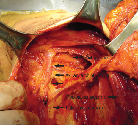
Fig 1 The Axillary arch of Langer in operation field (We identified an abnormal muscle originated from latissimus dorsi, and inserted to trilaminar tendon of pectoralis major during axillary lymph node dissection. The muscle interfered level I area due to its longitudinally oblique direction).
Reference
-
1. Miguel M, Llusá M, Ortiz JC, Porta N, Lorente M, Götzens V. The axillopectoral muscle (of Langer): report of three cases. Surg Radiol Anat. 2001. 23:341–343.
Article2. Boontje AH. Axillary vein entrapment. Br J Surg. 1979. 66:331–332.
Article3. Serpell JW, Baum M. Significance of 'Langer's axillary arch' in axillary dissection. Aust N Z J Surg. 1991. 61:310–312.
Article4. Daniels IR, della Rovere GQ. The axillary arch of Langer--the most common muscular variation in the axilla. Breast Cancer Res Treat. 2000. 59:77–80.
Article5. Dharap A. An unusually medial axillary arch muscle. J Anat. 1994. 184:639–641.6. Clarys JP, Barbaix E, Van Rompaey H, Caboor D, Van Roy P. The muscular arch of the axilla revisited: its possible role in the thoracic outlet and shoulder instability syndromes. Man Ther. 1996. 1:133–139.
Article7. Ko K, Han BK, Shin JH, Choe YH, Chung HW, Lee EH, et al. The axillopectoral muscle seen on mammography. Clin Radiol. 2006. 61:625–629.
Article8. Suzuma T, Sakurai T, Yoshimura G, Umemura T, Shimizu Y, Yang QF, et al. Magnetic resonance axillography for preoperative diagnosis of the axillopectoral muscle (Langer's axillary arch): a case report. Breast Cancer. 2003. 10:281–283.
Article9. Ucerler H, Ikiz ZA, Pinan Y. Clinical importance of the muscular arch of the axilla (axillopectoral muscle, Langer's axillary arch). Acta Chir Belg. 2005. 105:326–328.
Article10. Chêne G, Le Bouëdec G, Dauplat J. Arch and sentinel: surgical technique of sentinel node biopsy with the axillopectoral muscle. Gynecol Obstet Fertil. 2007. 35:25–29.11. Turgut HB, Peker T, Gülekon N, Anil A, Karaköse M. Axillopectoral muscle (Langer's muscle). Clin Anat. 2005. 18:220–223.
Article
- Full Text Links
-
- Actions
-
Cited
- CITED
-
- Close
- Share
-
- Similar articles
-
- Variation of the Axillary Arch in Korean Cadaver
- Duplicated axillary arch muscles arising from the latissimus dorsi
- A Comparison of Clinical Effect for Root Coverage
- Clinical Significance of the Axillary Arch in Sentinel Lymph Node Biopsy
- Short Rib Syndrome Beemer-Langer Type with Polydactyly: A Case Report
