A Comparative Study of Palpable and Nonpalpable Breast Cancers determined by Ultrasonography
- Affiliations
-
- 1Department of Radiology, Korea University Anam Hospital, Korea University College of Medicine, Seoul, Korea.
- 2Department of Radiology, Korea University Ansan Hospital, Korea University College of Medicine, Ansan, Korea. seoboky@korea.ac.kr
- 3Department of Biostatistics, Korea University College of Medicine, Seoul, Korea.
- 4Division of Oncology/Hematology, Department of Internal Medicine, Korea University Anam Hospital, Korea University College of Medicine, Seoul, Korea.
- 5Division of Oncology/Hematology, Department of Surgery, Korea University Anam Hospital, Korea University College of Medicine, Seoul, Korea.
- KMID: 2241933
- DOI: http://doi.org/10.4048/jbc.2008.11.2.64
Abstract
-
PURPOSE: The aim of this study was to investigate any difference of ultrasound findings for palpable and nonpalpable breast cancers.
METHODS
Two hundred breast cancer patients that had undergone preoperative ultrasound and surgery were enrolled in the study. A total of 126 cancers were palpable, and the remaining 74 cancers were nonpalpable. We compared lesion characteristics using ultrasound images according to the BI-RADS(R)-Ultrasound guidelines of the American College of Radiology. A crude odds ratio (OR) and 95% confidence interval (CI) were calculated for a comparison of the palpable and nonpalpable breast cancers.
RESULTS
Nonpalpable cancers displayed more often an oval shape (OR=0.35, 95% CI=0.17-0.70), no posterior acoustic features (OR=0.50, 95% CI=0.28-0.89), and a parallel orientation (OR=0.50, 95% CI=0.28-0.89). An irregular shape (OR=2.98, 95% CI=1.60-5.54), a spiculated margin (OR=2.66, 95% CI=1.23-5.74), and a combined pattern of posterior acoustic features (OR=7.20, 95% CI=1.64-31.66) were more commonly observed in the palpable cancers.
CONCLUSION
Palpable and nonpalpable breast cancers were found to have different ultrasound characteristics.
Keyword
Figure
-

Fig 1 64-yr-old woman with a palpable cancer. US shows a spiculated, irregular shaped, hypoechoic mass (arrows) that is not parallel to the skin, and which has a combined pattern of posterior acoustic feature. On pathologic examination, the mass proved to be a 12 mm sized invasive ductal carcinoma.
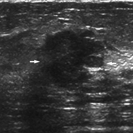
Fig 2 71-yr-old woman with a palpable cancer. US demonstrates a microlobulated, irregular shaped, hypoechoic mass (arrows) that is not parallel to the skin. This mass has a combined pattern of posterior acoustic feature. On pathologic examination, the mass proved to be a mixed invasive ductal and lobular carcinoma of size 22 mm.

Fig 3 48-yr-old woman with a nonpalpable cancer. US shows a microlobulated, oval, isoechoic mass (arrows) that is parallel to the skin. The mass has no posterior acoustic feature. On pathologic examination, the mass proved to be an invasive ductal carcinoma with low grade DCIS of size 6 mm.
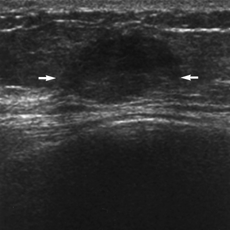
Fig 4 52-yr-old woman with a nonpalpable breast cancer. US shows a circumscribed, oval, hypoechoic mass (arrows) lying parallel to skin. On pathologic examination, the mass proved to be an invasive ductal carcinoma with high grade DCIS of size 12 mm.
Cited by 1 articles
-
Commentary on: Incidental Breast Cancers Identified in a One-Stop Symptomatic Breast Clinic
Jeong Eon Lee, Jung-Hyun Yang, Seok Jin Nam
J Breast Cancer. 2011;14(2):165-166. doi: 10.4048/jbc.2011.14.2.165.
Reference
-
1. Bassett LW, Liu TH, Giuliano AE, Gold RH. The prevalence of carcinoma in palpable vs impalpable, mammographically detected lesions. AJR Am J Roentgenol. 1991. 157:21–24.
Article2. Pagana TJ, Lubbe WJ, Schwartz SM, Sprechini GD. A comparison of palpable and nonpalpable breast cancers. Arch Surg. 1989. 124:26–28.
Article3. Perdue P, Page D, Nellestein M, Salem C, Galbo C, Ghosh B. Early detection of breast carcinoma: a comparison of palpable and nonpalpable lesions. Surgery. 1992. 111:656–659.4. Silverstein MJ, Gamagami P, Masetti R, Legmann MD, Craig PH, Gierson ED. Results from a multidisciplinary breast center. Analysis of disease discovered. Surg Oncol Clin N Am. 1997. 6:301–314.5. Silverstein MJ, Gierson ED, Waisman JR, Colburn WJ, Gamagami P. Predicting axillary node positivity in patients with invasive carcinoma of the breast by using a combination of T category and palpability. J Am Coll Surg. 1995. 180:700–704.6. Skinner KA, Silberman H, Sposto R, Silverstein MJ. Palpable breast cancers are inherently different from nonpalpable breast cancers. Ann Surg Oncol. 2001. 8:705–710.
Article7. Tafra L, Essner R, Brenner RJ, Giuliano AE. Nonpalpable versus palpable invasive breast tumors treated with breast-conserving surgical management. Am Surg. 1996. 62:395–399.8. Burrell HC, Sibbering DM, Wilson AR, Pinder SE, Evans AJ, Yeoman LJ, et al. Screening interval breast cancers: mammographic features and prognosis factors. Radiology. 1996. 199:811–817.
Article9. Hollingsworth AB, Taylor LD, Rhodes DC. Establishing a histologic basis for false-negative mammograms. Am J Surg. 1993. 166:643–647.
Article10. Laya MB, Larson EB, Taplin SH, White E. Effect of estrogen replacement therapy on the specificity and sensitivity of screening mammography. J Natl Cancer Inst. 1996. 88:643–649.
Article11. Kerlikowske K, Grady D, Barclay J, Sickles EA, Ernster V. Effect of age, breast density, and family history on the sensitivity of first screening mammography. JAMA. 1996. 276:33–38.
Article12. Oza AM, Boyd NF. Mammographic parenchymal patterns: a marker of breast cancer risk. Epidemiol Rev. 1993. 15:196–208.
Article13. Saarenmaa I, Salminen T, Geiger U, Heikkinen P, Hyvarinen S, Isola J, et al. The effect of age and density of the breast on the sensitivity of breast cancer diagnostic by mammography and ultasonography. Breast Cancer Res Treat. 2001. 67:117–123.
Article14. Saftlas AF, Wolfe JN, Hoover RN, Brinton LA, Schairer C, Salane M, et al. Mammographic parenchymal patterns as indicators of breast cancer risk. Am J Epidemiol. 1989. 129:518–526.
Article15. Berg WA, Gilbreath PL. Multicentric and multifocal cancer: wholebreast US in preoperative evaluation. Radiology. 2000. 214:59–66.
Article16. Buchberger W, DeKoekkoek-Doll P, Springer P, Obrist P, Dunser M. Incidental findings on sonography of the breast: clinical significance and diagnostic workup. AJR Am J Roentgenol. 1999. 173:921–927.
Article17. Gordon PB, Goldenberg SL. Malignant breast masses detected only by ultrasound. A retrospective review. Cancer. 1995. 76:626–630.
Article18. Crystal P, Strano SD, Shcharynski S, Koretz MJ. Using sonography to screen women with mammographically dense breasts. AJR Am J Roentgenol. 2003. 181:177–182.
Article19. Kaplan SS. Clinical utility of bilateral whole-breast US in the evaluation of women with dense breast tissue. Radiology. 2001. 221:641–649.
Article20. Kolb TM, Lichy J, Newhouse JH. Occult cancer in women with dense breasts: detection with screening US--diagnostic yield and tumor characteristics. Radiology. 1998. 207:191–199.
Article21. Leconte I, Feger C, Galant C, Berliere M, Berg BV, D'Hoore W, et al. Mammography and subsequent whole-breast sonography of nonpalpable breast cancers: the importance of radiologic breast density. AJR Am J Roentgenol. 2003. 180:1675–1679.
Article22. Stavros AT, Thickman D, Rapp CL, Dennis MA, Parker SH, Sisney GA. Solid breast nodules: use of sonography to distinguish between benign and malignant lesions. Radiology. 1995. 196:123–134.
Article23. American College of Radiology. BI-RADS Committee. . Breast ultrasound. ACR BI-RADS breast imaging and reporting data system: breast imaging atlas. 2003. (1st ed). Reston, VA: American College of Radiology.24. Thurfjell MG, Lindgren A, Thurfjell E. Nonpalpable breast cancer: mammographic appearance as predictor of histologic type. Radiology. 2002. 222:165–170.
Article25. Lamb PM, Perry NM, Vinnicombe SJ, Wells CA. Correlation between ultrasound characteristics, mammographic findings and histological grade in patients with invasive ductal carcinoma of the breast. Clin Radiol. 2000. 55:40–44.
Article26. Moon WK, Myung JS, Lee YJ, Park IA, Noh DY, Im JG. US of ductal carcinoma in situ. Radiographics. 2002. 22:269–280.
Article27. Colpaert C, Vermeulen P, van Beest P, Goovaerts G, Weyler J, Van Dam P, et al. Intratumoral hypoxia resulting in the presence of a fibrotic focus is an independent predictor of early distant relapse in lymph node-negative breast cancer patients. Histopathology. 2001. 39:416–425.
Article28. Chiou SY, Chou YH, Chiou HJ, Wang HK, Tiu CM, Tseng LM, et al. Sonographic features of nonpalpable breast cancer: a study based on ultrasound-guided wire-localized surgical biopsies. Ultrasound Med Biol. 2006. 32:1299–1306.
Article
- Full Text Links
-
- Actions
-
Cited
- CITED
-
- Close
- Share
-
- Similar articles
-
- The Clinicopathological Characteristics of Palpable and Non-palpable Breast Cancer
- Evaluation of diagnostic methods in children with nonpalpable undescended testis
- Comparison between Mammography and Ultrasonography for Palpable Breast Mass: Focusing Fibroadenoma and Breast Cancer
- Localization of Nonpalpable Breast lesion with Ultrasonoguided Dye Injection
- Needle Localization Biopsy of Nonpalpable lesions of Breast under the Local Anesthesia
