Superficial Fibromatosis Mimicking Glomus Tumor of the Second Toe
- Affiliations
-
- 1Department of Pathology, Gunsan Medical Center of Wonkwang University Hospital, Gunsan, Korea.
- 2Department of Orthopedic Surgery, Gunsan Medical Center of Wonkwang University Hospital, Gunsan, Korea. oschae68@hanmail.net
- 3Department of Radiology, Gunsan Medical Center of Wonkwang University Hospital, Gunsan, Korea.
- KMID: 2234102
- DOI: http://doi.org/10.4055/cios.2015.7.3.418
Abstract
- Various types of tumor can occur in the subungual space, including glomus tumors, subungual exostosis, hemangioma, epidermal cysts, and malignant tumors. While fibromatosis can occur at various sites throughout the body, it is very rarely seen in the toe. Here, we are the first to report a case of superficial fibromatosis mimicking a glomus tumor in the subungual space of the second toe. The presentation of this condition shows the possibility of encountering uncommon superficial fibromatosis in the distal phalanx of the toe, and suggests that superficial fibromatosis should be included in the differential diagnosis of a glomus tumor in the toe.
Keyword
Figure
-
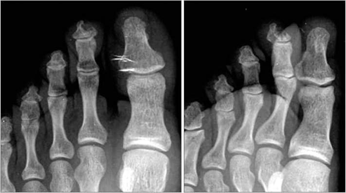
Fig. 1 Radiographs of the left foot showing the focal osteolytic lesion with a subtle sclerotic margin in the distal phalanx of second toe.
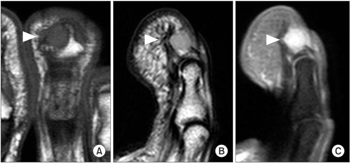
Fig. 2 Coronal T1-weighted (A), sagittal T2-weighted (B), and sagittal gadolinium-enhanced fat-saturated T1-weighted (C) magnetic resonance imaging scans of the left foot showing the approximate 0.7 × 0.6 × 0.5 cm well-marginated osteolytic lesion and low-signal rim with iso signal intensity on T1-weighted and high signal intensity on T2-weighted images compared to adjacent muscles, and homogeneous contrast enhancement in the distal phalanx of the second toe (arrowheads).
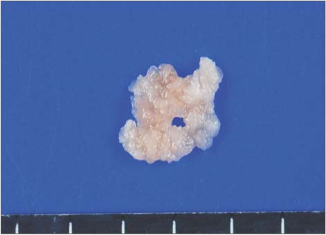
Fig. 3 The gross specimen shows a grayish white soft tissue.
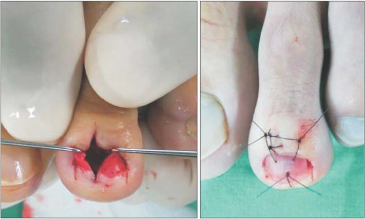
Fig. 4 Intraoperative photographs showing the nail bed longitudinal incision and reattachment of the removed nail as a nail bed protector.
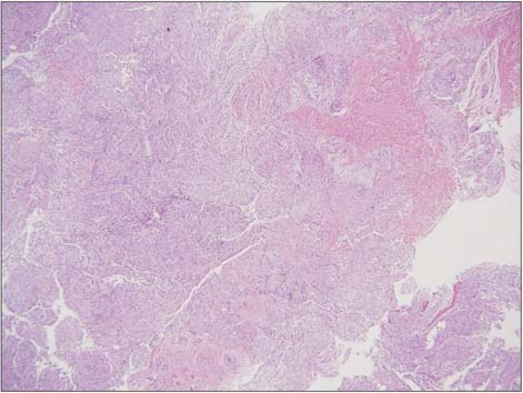
Fig. 5 The tumor shows cellular proliferation of bland spindled cells arranged into ill-defined long fascicles (H&E, ×40).

Fig. 6 The tumor cells are spindle cells without nuclear atypia (H&E, ×400).
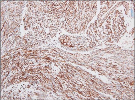
Fig. 7 The tumor cells are diffusely immunoreactive for vimentin (immunohistochemical stains, ×200).
Reference
-
1. Allen PJ, Shriver CD. Desmoid tumors of the chest wall. Semin Thorac Cardiovasc Surg. 1999; 11(3):264–269.
Article2. Pater TJ, Marks RM. Glomus tumor of the hallux: case presentation and review of the literature. Foot Ankle Int. 2004; 25(6):434–437.
Article3. Drape JL, Idy-Peretti I, Goettmann S, Guerin-Surville H, Bittoun J. Standard and high resolution magnetic resonance imaging of glomus tumors of toes and fingertips. J Am Acad Dermatol. 1996; 35(4):550–555.
Article4. Matloub HS, Muoneke VN, Prevel CD, Sanger JR, Yousif NJ. Glomus tumor imaging: use of MRI for localization of occult lesions. J Hand Surg Am. 1992; 17(3):472–475.
Article5. Baek HJ, Lee SJ, Cho KH, et al. Subungual tumors: clinicopathologic correlation with US and MR imaging findings. Radiographics. 2010; 30(6):1621–1636.
Article6. Guglielmi G, Cifaratti A, Scalzo G, Magarelli N. Imaging of superficial and deep fibromatosis. Radiol Med. 2009; 114(8):1292–1307.
Article7. Rosai J. Rosai and Ackerman's surgical pathology. 9th ed. Edinburgh: Mosby;2004.8. Robbin MR, Murphey MD, Temple HT, Kransdorf MJ, Choi JJ. Imaging of musculoskeletal fibromatosis. Radiographics. 2001; 21(3):585–600.
Article9. Mehrotra AK, Sheikh S, Aaron AD, Montgomery E, Goldblum JR. Fibromatoses of the extremities: clinicopathologic study of 36 cases. J Surg Oncol. 2000; 74(4):291–296.
Article10. Carroll RE. Juvenile aponeurotic fibroma. Hand Clin. 1987; 3(2):219–224.
Article
