Anomalous Origin of a Right Coronary Artery With Extrinsic Compression Between the Great Vessels: The Intravascular Ultrasound Images
- Affiliations
-
- 1Cardiovascular Research Institute of Chonnam National University, The Heart Center of Chonnam National University Hospital, Gwangju, Korea. myungho@chollian.net
- KMID: 2225774
- DOI: http://doi.org/10.4070/kcj.2008.38.7.390
Abstract
- The anomalous origin of the right coronary artery is a rare condition, but it has clinical importance because there have been reports of nonfatal or fatal myocardial infarction and sudden death associated with exercise for patients with this anatomy. We describe here a patient for whom 64 channel multi-detector row computed tomography was useful to identify this anomaly, and intravascular ultrasound was used to evaluate the myocardial ischemia by visualizing the coronary lumen.
MeSH Terms
Figure
-
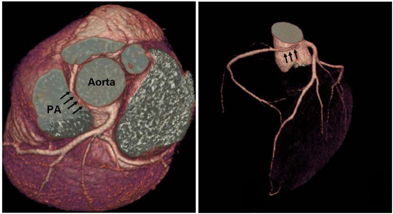
Fig. 1 Sixty four-slice CT image on the coronary artery. The RCA originated from the left coronary cusp with subsequent extrinsic compression between the aorta and pulmonary artery. RCA: right coronary artery, PA: pulmonary artery.
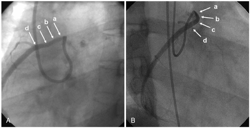
Fig. 2 Conventional coronary angiogram of the RCA. There was no critical stenotic lesion on the LAO 45° view (A). Critical stenosis was noted on the RAO 45° image (B) (the IVUS images of a, b, c and d are presented in Fig. 3). RCA: right coronary artery, LAO: left anterior oblique, RAO: right anterior oblique, IVUS: intravascular ultrasound.
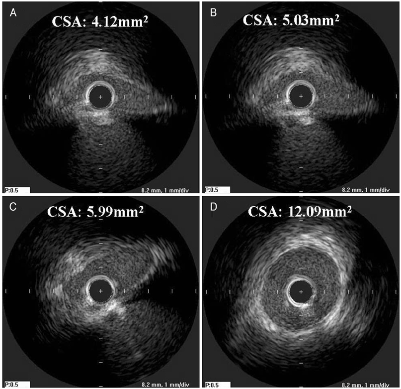
Fig. 3 Intravascular ultrasound (IVUS) of the RCA. The IVUS study showed no atherosclerotic plaque burdens on the entire RCA. However, the lumen was squeezed to a spindle shape (A) in the proximal portions because of the extrinsic compression between two large vessels (angiographic images of A, B, C and D are presented in Fig. 2). RCA: right coronary artery, CSA: cross sectional area.
Reference
-
1. Yamanaka O, Hobbs RE. Coronary artery anomalies in 126,595 patients undergoing coronary arteriography. Cathet Cardiovasc Diagn. 1990. 21:28–40.2. Roberts WC, Siegel RJ, Zipes DP. Origin of the right coronary artery from the left sinus of valsalva and its functional consequences: analysis of 10 necropsy patients. Am J Cardiol. 1982. 49:863–868.3. Kragel AH, Roberts WC. Anomalous origin of either the right or left main coronary artery from the aorta with subsequent coursing between aorta and pulmonary trunk: analysis of 32 necropsy cases. Am J Cardiol. 1988. 62:771–777.4. Ichikawa M, Sato Y, Komatsu S, Hirayama A, Kodama K, Saito S. Multislice computed tomographic findings of the anomalous origins of the right coronary artery: evaluation of possible causes of myocardial ischemia. Int J Cardiovasc Imaging. 2007. 23:353–360.5. Sato Y, Inoue F, Kunimasa T, et al. Diagnosis of anomalous origin of the right coronary artery using multislice computed tomography: evaluation of possible causes of myocardial ischemia. Heart Vessels. 2005. 20:298–300.6. Benge W, Martins JB, Funk DC. Morbidity associated with anomalous origin of the right coronary artery from the left sinus of valsalva. Am Heart J. 1980. 99:96–100.7. Virmani R, Chun PK, Goldstein RE, Robinowitz M, McAllister HA. Acute takeoffs of the coronary arteries along the aortic wall and congenital coronary ostial valve-like ridges: association with sudden death. J Am Coll Cardiol. 1984. 3:766–771.8. Kim MS, Han JK, Lee SE, et al. Cases of right ventricular myocardial infarction in patients with an absent or hypoplastic right coronary artery. Korean Circ J. 2007. 37:84–86.9. Shin SH, Park CG, Kim YH, et al. Anomalous origin of the right coronary artery from the diagonal branch of the left anterior descending coronary artery. Korean Circ J. 2004. 34:615–617.10. Hyun DW, Hur SH, Han SW. A case of right coronary artery originating from distal left circumflex (single coronary artery). Korean Circ J. 2003. 33:1044–1047.
- Full Text Links
-
- Actions
-
Cited
- CITED
-
- Close
- Share
-
- Similar articles
-
- Two Cases of Anomalous Origin of Coronary Artery
- Anomalous origin of the left coronary artery from the pulmonary artery
- Cardiopulmonary bypass weaning difficulty due to anomalous origin of coronary artery: A case report
- Sudden Death Associated with Anomalous Left Coronary Artery Origin from Right Sinus of Valsalva with Posterior Course
- Anomalous Origin of Left Main Coronary Artery from the Right Coronary Artery: Echocardiographic Diagnosis
