Korean Circ J.
2008 Oct;38(10):551-556. 10.4070/kcj.2008.38.10.551.
Relationship Between RR Intervals and Early Diastolic Mitral Annulus Velocities in Atrial Fibrillation Patients Who do not Have Significant Valvular Diseases
- Affiliations
-
- 1Division of Cardiology, Department of Internal Medicine, College of Medicine, Chung-Ang University, Seoul, Korea. cjkim@cau.ac.kr
- KMID: 2225720
- DOI: http://doi.org/10.4070/kcj.2008.38.10.551
Abstract
- BACKGROUND AND OBJECTIVES
Irregular RR intervals in atrial fibrillation (AF) results in beat to beat changes in hemodynamical parameters. Early diastolic mitral annulus velocity (E') is one of the parameters that represent diastolic function of the left ventricle (LV). In this study, we have investigated the effects of continuous changes of systolic functions in AF on the diastolic functions of the LV. SUBJECTS AND METHODS: E' (35-40 beats) was recorded in 31 AF patients that did not have significant valvular heart diseases. The relationships between preceding RR intervals (RR-1) or pre-preceding RR intervals (RR-2) and E's were obtained using a logarithmic function. RESULTS: Slopes between RR-1 and E' varied from -1.62 to 1.04 in total coordinates. In the logistic regression analysis patients with negative slopes were found to have a larger left atrial size than patients with positive slopes (5.5+/-0.67 cm vs. 4.9+/-0.56 cm, p=0.02). Slopes were negatively related with mean RR intervals in the Pearson correlation analysis (r=-0.40, p=0.028). Slopes between RR-2 and E' were also variable and were not associated with other parameters. CONCLUSION: Beat to beat changes in systolic functions derived from irregular RR intervals in AF had variable effects on diastolic functions among patients. The relationship between RR-1 and E' was associated with LA sizes and mean RR intervals.
Keyword
MeSH Terms
Figure
-
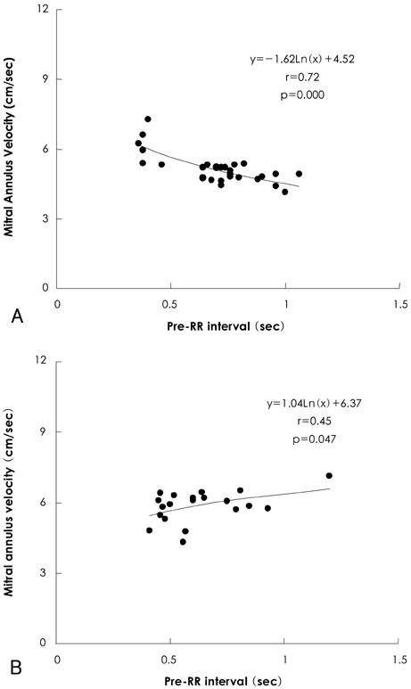
Fig. 1 Representative examples of negative (A) and positive (B) relationships between preceding RR intervals and early diastolic mitral annulus velocities.
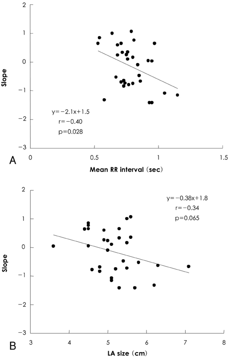
Fig. 2 Relationship of slopes between preceding RR intervals and early diastolic mitral annulus velocities with mean RR intervals (A) and left atrial (LA) sizes (B).
Reference
-
1. Sohn DW, Chai IH, Lee DJ, et al. Assessment of mitral annulus velocity by Doppler tissue imaging in the evaluation of left ventricular diastolic function. J Am Coll Cardiol. 1997. 30:474–480.2. Ha JW, Oh JK. The pathophysiology and diagnostic approaches for diastolic left ventricular dysfunction: a clinical perspective. Korean Circ J. 2005. 35:865–876.3. Kim KS. The usefulness of Doppler tissue image in evaluation of left ventricular systolic and diastolic dysfunction. Korean Circ J. 2002. 32:99–105.4. De Boeck BW, Cramer MJ, Oh JK, van der Aa RP, Jaarsma W. Spectral pulsed tissue Doppler imaging in diastole: a tool to increase our insight in and assessment of diastolic relaxation of the left ventricle. Am Heart J. 2003. 146:411–419.5. Oki T, Tabata T, Yamada H, et al. Clinical application of pulsed Doppler tissue imaging for assessing abnormal left ventricular relaxation. Am J Cardiol. 1997. 79:921–928.6. Nagueh SF, Middleton KJ, Kopelen HA, Zoghbi WA, Quiñones MA. Doppler tissue imaging: a noninvasive technique for evaluation of left ventricular relaxation and estimation of filling pressures. J Am Coll Cardiol. 1997. 30:1527–1533.7. Gosselink AT, Blanksma PK, Crijns HJ, et al. Left ventricular beat-to-beat performance in atrial fibrillation: contribution of Frank-Starling mechanism after short rather than long RR intervals. J Am Coll Cardiol. 1995. 26:1516–1521.8. Freeman GL, Colston JT. Evaluation of left ventricular mechanical restitution in closed-chest dogs based on single-beat elastance. Circ Res. 1990. 67:1437–1445.9. Cho SW, Kim SM, Lee SY, et al. The relationship between preceding RR intervals and peak ejection velocity of left ventricular outflow in atrial fibrillation without organic heart diseases. Korean Circ J. 2003. 33:113–120.10. Ko HS, Lee KJ, Kim SW, Kim TH, Kim CJ, Ryu WS. Prediction of left ventricular peak ejection velocity by preceding and prepreceding RR intervals in atrial fibrillation: a new method to adjust the influence between two Intervals. J Korean Med Sci. 2002. 17:743–748.11. Sahn DJ, DeMaria A, Kisslo J, Weyman A. Recommendations regarding quantitation in M-mode echocardiography: results of a survey of echocardiographic measurements. Circulation. 1978. 58:1072–1083.12. Aranda JM Jr, Weston MW, Puleo JA, Fontanet HL. Effect of loading conditions on myocardial relaxation velocities determined by Doppler tissue imaging in heart transplant recipients. J Heart Lung Transplant. 1998. 17:693–697.13. Puleo JA, Aranda JM, Weston MW, et al. Noninvasive detection of allograft rejection in heart transplant recipients by use of Doppler tissue imaging. J Heart Lung Transplant. 1998. 17:176–184.14. Nagueh SF, Kopelen HA, Quiñones MA. Assessment of left ventricular filling pressures by Doppler in the presence of atrial fibrillation. Circulation. 1996. 94:2138–2145.15. Ommen SR, Nishimura RA, Appleton CP, et al. Clinical utility of Doppler echocardiography and tissue Doppler imaging in the estimation of left ventricular filling pressures: a comparative simultaneous Doppler-catheterization study. Circulation. 2000. 102:1788–1794.16. Sohn DW, Song JM, Zo JH, et al. Mitral annulus velocity in the evaluation of left ventricular diastolic function in atrial fibrillation. J Am Soc Echocardiogr. 1999. 12:927–931.17. Chirillo F, Brunazzi MC, Barbiero M, et al. Estimating mean pulmonary wedge pressure in patients with chronic atrial fibrillation from transthoracic Doppler indexes of mitral and pulmonary venous flow velocity. J Am Coll Cardiol. 1997. 30:19–26.18. Yamada H, Oki T, Mishiro Y, et al. Effect of aging on diastolic left ventricular myocardial velocities measured by pulsed tissue Doppler imaging in healthy subjects. J Am Soc Echocardiogr. 1999. 12:574–581.
- Full Text Links
-
- Actions
-
Cited
- CITED
-
- Close
- Share
-
- Similar articles
-
- Relation Between RR Intervals and Early Diastolic Mitral Annular Velocities in Atrial Fibrillation
- Relation between Left Atrial Size and Atrial Fibrillation
- Assessment of Mitral Valve Area in Patients with Mitral Stenosis and Atrial Fibrillation
- Relation of Right Atrial Pathology to Atrial Fibrillation in Mitral Valvular Disease
- Echocardiographic Study on the Mitral Valvular Heart Diseases
