How Does the Left Ventricle Work? Ventricular Rotation as a New Index of Cardiac Performance
- Affiliations
-
- 1Division of Cardiology, Asan Medical Center, University of Ulsan College of Medicine, Seoul, Korea. jksong@amc.seoul.kr
- KMID: 2225669
- DOI: http://doi.org/10.4070/kcj.2009.39.9.347
Abstract
- Although simple cylindrical or ellipsoidal left ventricular (LV) geometry with transverse or circumferential muscle contraction has been traditionally used to estimate LV performance, the estimated LV ejection fraction (EF) with muscle fiber shortening up to 20% is less than 50% of maximum, which is lower than the normal EF observed in routine clinical practice. Thus, oblique fiber orientation and LV rotation, in addition to radial thickening and longitudinal shortening, is predicted as an essential component of effective LV pumping. This was confirmed by animal experiments using surgically implanted markers or invasive sonomicrometry. Demonstration of the muscle band extending from the pulmonary artery to the aorta, which connects the ventricular myocardium, both right ventricle and LV as a continuous band (muscle band theory) provides an anatomical backbone of helical configuration of the cardiac muscle band with descending and ascending segments wrapping the LV apex. Moreover, sequential, non-simultaneous, activation and contraction of the helicoids muscle band contributes to LV rotation or twist motion. Recently, magnetic resonance imaging and speckle tracking echocardiography (STE) techniques have provided an excellent noninvasive way to measure LV rotation and twist, which is expected to contribute to a more thorough evaluation of both LV systolic and diastolic function. Initial animal experiments showed that quantification of apical rotation or LV twist using STE is more accurate for estimating LV systolic function than conventional EF under a variety of LV inotropic conditions, irrespective of coronary ligation. As de-rotation or the untwisting rate can also be measured by STE, the role of ventricular untwisting as a temporal link between LV relaxation and suction can be addressed. Further clinical investigations are needed to determine the real clinical impact of these new indices of LV mechanical function.
Keyword
MeSH Terms
Figure
-

Fig. 1 Diagrams showing various fiber geometries and mathematical models of the left ventricle for calculation of ejection fraction (EF). Upon stimulation, uniform shortening of the individual fibers to k times their initial lengths results in a radius change from B to kB with the indicated EF. If k=0.8 in circular fibers, which corresponds to a 20% shortening, this corresponds to an EF of only 0.36 for the cylinder (A) or ellipse of revolution (B) and 0.488 for the sphere (C). In a cylindrical model with longitudinal fibers (D), 15% fiber shortening results in only a 15% reduction in chamber volume, whereas with circumferential fibers (E), 15% fiber shortening results in only a 30% reduction in volume. Depending only upon the cylinder dimensions and the pitch angle of the spiral fibers (F), virtually any ejection fraction (up to and including 100%) could be generated from spiral fibers capable of shortening only 15% (modified from references 1 and 2).
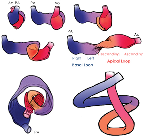
Fig. 2 Representative illustrations demonstrating the muscle band extending from the pulmonary artery (PA) to the aorta (Ao) which connects the ventricular myocardium, both right ventricle and LV, as a continuous band. This band provides an anatomical backbone of helical configuration of the cardiac muscle band with descending and ascending segments wrapping the LV apex. The helical configuration of the cardiac muscle band results in two loops at the cardiac base and apex (modified from reference 5). LV: left ventricular.

Fig. 3 Representative images of apical and basal rotation angles with corresponding rotational velocity curves measured by speckle tracking echocardiography.
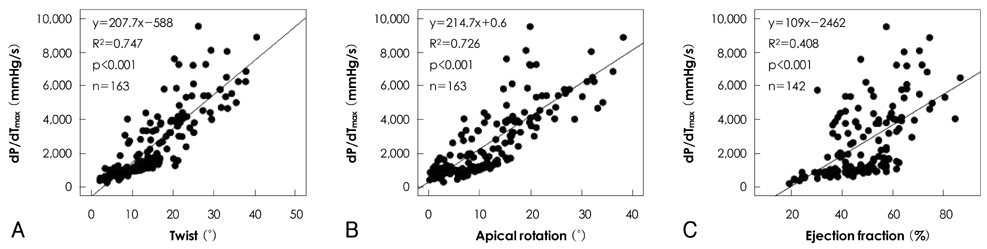
Fig. 4 Association between dP/dtmax, an invasive gold standard of LV contractility and left ventricular twist (A), apical rotation (B) and ejection fraction (C)(modified from reference 15). LV: left ventricular.
Reference
-
1. Sallin EA. Fiber orientation and ejection fraction in the human left ventricle. Biophys J. 1969. 9:954–964.2. Ingels NB Jr. Myocardial fiber architecture and left ventricular function. Technol Health Care. 1997. 5:45–52.3. Lower R. Gunther RT, editor. Tractus de corde. Early Science in Oxford. 1968. Reprint. Oxford, UK: Dawsons, Pall Mall, London;1669.4. Ingels NB, Daughters GT 2nd, Stinson EB, Alderman EL. Measurement of midwall myocardial dynamics in intact man by radiography of surgically implanted markers. Circulation. 1975. 52:859–867.5. Torrent-Guasp F, Ballester M, Buckberg GD, et al. Spatial orientation of the ventricular muscle band: physiologic contribution and surgical implications. J Thorac Cardiovasc Surg. 2001. 122:389–392.6. Prinzen FW, Augustijn CH, Allessie MA, Arts T, Delhaas T, Reneman RS. The time sequence of electrical and mechanical activation during spontaneous beating and ectopic stimulation. Eur Heart J. 1992. 13:535–543.7. Helm RH, Lecercq C, Faris OP, et al. Cardiac dyssynchrony analysis using circumferential versus longitudian strain: implications for assessing cardiac resynchronization. Circulation. 2005. 111:2760–2767.8. Smiseth OA, Remme EW. Regional left ventricular electric and mechanical activation and relaxation. J Am Coll Cardiol. 2006. 47:173–174.9. Sengupta PP, Korinek J, Belohlavek M, et al. Left ventricular structure and function: basic science for cardiac imaging. J Am Coll Cardiol. 2006. 48:1988–2001.10. Tomioka H, Liakopoulos OJ, Buckberg GD, Hristov N, Tan Z, Trummer G. The effect of ventricular sequential contraction on helical heart during pacing: high septal pacing versus biventricular pacing. Eur J Cardiothoracic Surg. 2006. 29:Suppl 1. S198–S206.11. Hansen DE, Daughters GT 2nd, Alderman EL, Ingels NB, Stinson EB, Miller DC. Effect of volume loading, pressure loading, and inotropic stimulation on left ventricular torsion in humans. Circulation. 1991. 83:1315–1326.12. Moon MR, Ingels NB Jr, Daughters GT 2nd, Sinson EB, Hansen DE, Miller DC. Alterations in left ventricular twist mechanics with inotropic stimulation and volume loading in human subjects. Circulation. 1994. 89:142–150.13. Buchalter MB, Rademakers FE, Weiss JL, Rogers WJ, Weisfeldt ML, Shapiro EP. Rotational deformation of the canine left ventricle measured by magnetic resonance tagging: effects of catecholamines, ischemia, and pacing. Cardiovasc Res. 1994. 28:629–635.14. Kroeker CA, Tyberg JV, Beyar R. Effects of on left ventricular apical rotation: an experimental study in anesthetized dogs. Circulation. 1995. 92:3539–3548.15. Kim WJ, Lee BH, Kim YJ, et al. Apical rotation assessed by speckle-tracking echocardiography as an index of global left ventricular contractility. Circ Cardiovasc Imaging. 2009. 2:123–131.16. Kim HK, Sohn DW, Lee SE, et al. Assessment of left ventricular rotation and torsion with two-dimensional speckle tracking echocardiography. J Am Soc Echocardiogr. 2007. 20:45–53.17. Notomi Y, Popovic ZB, Yamada H, et al. Ventricular untwisting: a temporal link between left ventricular relaxation and suction. Am J Physiol Heart Circ Physiol. 2007. 294:H505–H513.
- Full Text Links
-
- Actions
-
Cited
- CITED
-
- Close
- Share
-
- Similar articles
-
- Left Ventricular Rotation and Twist: Why Should We Learn?
- Relation of hemodynamic load to left ventricular hypertrophy and performance in essential hypertension
- Double Outlet of Right Ventricle in Criss-Cross Heart: Surgical Experience of One Case
- Comparison of Left and Right Ventricular Volume and Cardiac Output by MRI and Echocardiography
- Aneurysm or Diverticulum of Left Ventricle
