Korean Circ J.
2010 Jul;40(7):352-353. 10.4070/kcj.2010.40.7.352.
Intracardiac Tumor Presenting as Complete Atrioventricular Block
- Affiliations
-
- 1Department of Cardiology, Sunlin Hospital, Pohang, Korea. andesdr@yahoo.com
- 2Division of Cardiology, Department of Internal Medicine, College of Medicine, Inje University, Paik Hospital, Busan, Korea.
- KMID: 2225181
- DOI: http://doi.org/10.4070/kcj.2010.40.7.352
Abstract
- No abstract available.
MeSH Terms
Figure
-
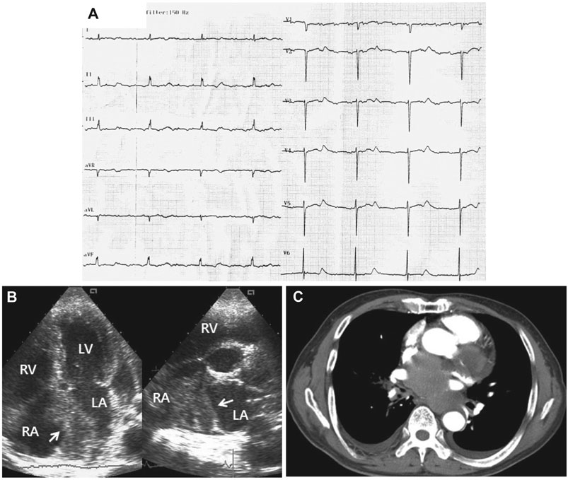
Fig. 1 An electrocardiogram showing sinus tachycardia with complete AV block (A). An echolucent inhomogenous mass (arrows) involving the interatrial septum and protruding toward both atria was found on the echocardiogram (B). Chest computed tomography revealed a large non-enhanced hypodense mass invading the atria, the ventricles, and the pulmonary trunk (C). AV: atrioventricular.
Reference
-
1. Min DJ, Kang DH, Seung KB, et al. A primary cardiac angiosarcoma. Korean Circ J. 1995. 25:704–709.2. Kwon YJ, Seo SW, Kim SG. A case of primary fibrosarcoma in left atrium. Korean Circ J. 1987. 17:389–393.3. Castilla-Cabanes E, Pascual-Calleja I, Roncales-Garcia Blanco F, del Rio-Ligorit A. Clinical variations of cardiac sarcoma. Rev Esp Cardiol. 2009. 62:823–824.4. Lurito KJ, Martin T, Cordes T. Right atrial primary cardiac osteosarcoma. Pediatr Cardiol. 2002. 23:462–465.5. Lee GC, Cho JG, Choi SJ, et al. A case of malignant lymphoma with cardiac involvement at initial presentation. Korean Circ J. 1994. 24:899–903.
- Full Text Links
-
- Actions
-
Cited
- CITED
-
- Close
- Share
-
- Similar articles
-
- A case of complete atrioventricular block persisting for 5 days in a patient with variant angina
- A Case of Transient Complete Atrioventricular Block in Acute Viral Myocarditis
- A Case of Hyperthyroidism with Complete Atrioventricular Block and Cardiac Arrest
- A Case of Fetal Complete Atrioventricular Block Corrected by Cardiac Pacemaker Implantation after Birth
- Atrioventricular nodal re‑entrant tachycardia with a 2:1 atrioventricular block in a young man: What is the mechanism?
