Rupture and Spontaneous Sealing of a Coronary Aneurysm After Deployment of Drug-Eluting Stent
- Affiliations
-
- 1Department of Internal Medicine, Gyeongsang National University Hospital, Jinju, Korea. jyhwang@nongae.gsnu.ac.kr
- KMID: 2225006
- DOI: http://doi.org/10.4070/kcj.2012.42.8.558
Abstract
- Lesions with coronary artery aneurysm (CAA) can become complicated during percutaneous coronary intervention. Here, we report a case of a 78-year-old man who developed a rupture, and spontaneous sealing of the CAA occurred after stent implantation, as shown by computed tomography coronary angiography.
Keyword
MeSH Terms
Figure
-
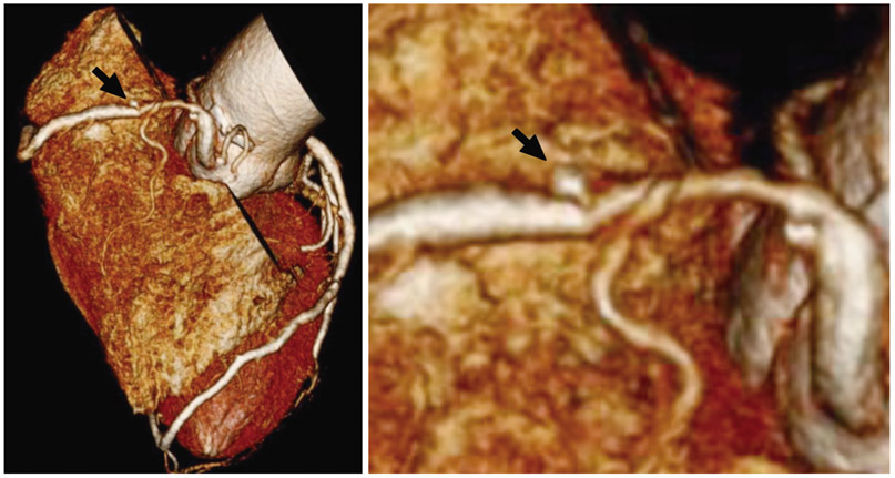
Fig. 1 Baseline three-dimensional reconstruction image of computed tomography coronary angiography. A three-dimensional reconstruction image of multidetector computed tomography coronary angiography shows a 2.5×2.5 mm saccular coronary aneurysm (black arrow) and tubular stenosis (60-70%) on the proximal right coronary artery.
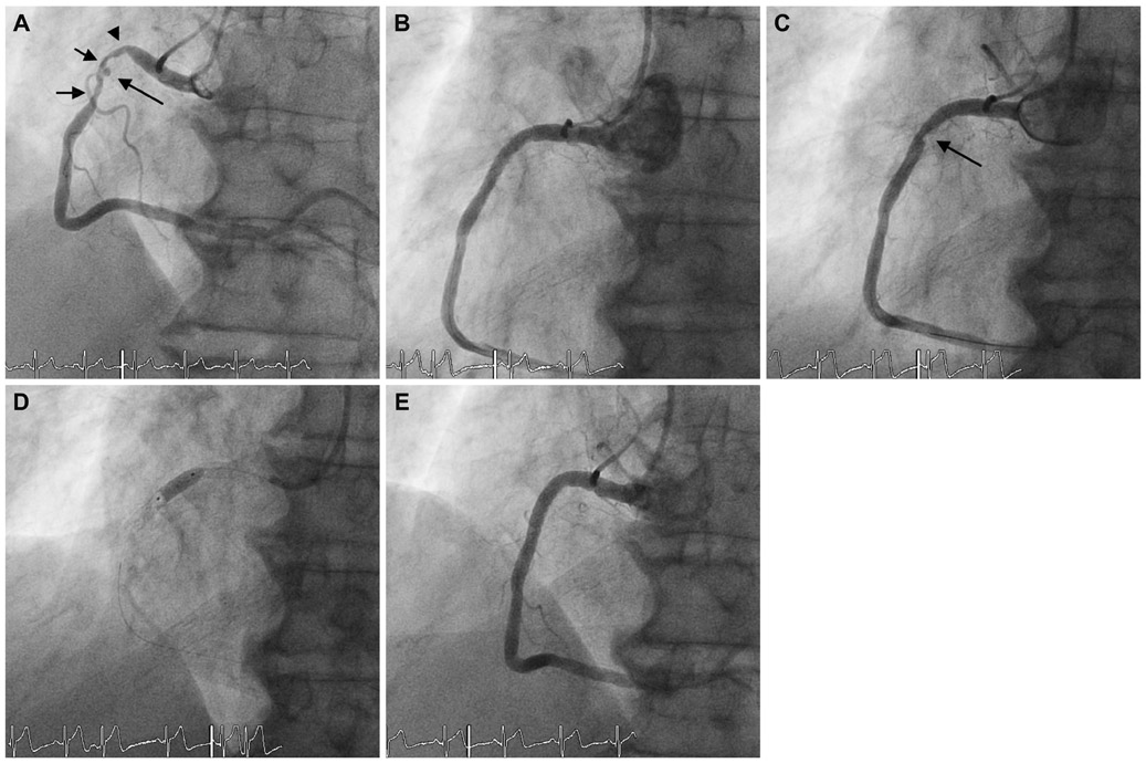
Fig. 2 Coronary angiography of the right coronary artery. A: coronary angiography shows a saccular coronary artery aneurysm (large arrow) with diffuse, concentric stenosis in the proximal portion of the RCA (arrowhead). After stenting, the coronary aneurysm was no longer seen, and 1 side branch of the proximal RCA was jailed (arrow). B: angiography shows thrombolysis in myocardial infarction 3 flow and no luminal defect. C: after adjunctive ballooning, the thrombus is visualized inside the stent by the lack of contrast filling (arrow). D: balloon dilatation is performed at the site of the thrombus. E: after a second stent deployment, any filling defect suggestive of thrombus formation disappeared and good angiographic results were observed. RCA: right coronary artery.
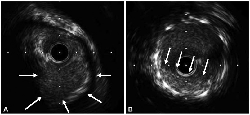
Fig. 3 Intravacular ultrasound image. A: intravascular ultrasonography shows 2.5×2.5 mm sized small aneurysms (arrows) and fibro-fatty plaque in the proximal right coronary artery. B: after stenting, the intravascular ultrasound image shows a thrombus inside the stent (arrows), but no aneurysms were seen.
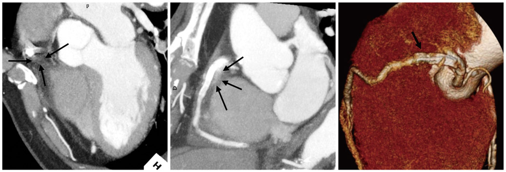
Fig. 4 CT coronary angiography after coronary intervention. CT coronary angiography 10 days after coronary intervention shows a ruptured and sealed coronary aneurysm along the proximal right coronary artery (arrow).
Reference
-
1. Syed M, Lesch M. Coronary artery aneurysm: a review. Prog Cardiovasc Dis. 1997. 40:77–84.2. Vijayanagar R, Shafii E, DeSantis M, Waters RS, Desai A. Surgical treatment of coronary aneurysms with and without rupture. J Thorac Cardiovasc Surg. 1994. 107:1532–1535.3. Cohen P, O'Gara PT. Coronary artery aneurysms: a review of the natural history, pathophysiology, and management. Cardiol Rev. 2008. 16:301–304.4. Hayat SA, Ghani S, More RS. Treatment of ruptured coronary aneurysm with a novel covered stent. Catheter Cardiovasc Interv. 2009. 74:367–370.5. Alford WC Jr, Stoney WS, Burrus GR, Frist RA, Thomas CS Jr. Recognition and operative management of patients with arteriosclerotic coronary artery aneurysms. Ann Thorac Surg. 1976. 22:317–321.6. Kim HG, Jeong MH, Kim W, et al. Successful stent grafting for a coronary aneurysm. Korean Circ J. 2004. 34:507–511.7. Gunduz H, Akdemir R, Binak E, Tamer A, Uyan C. Spontaneous rupture of a coronary artery aneurysm: a case report and review of the literature. Jpn Heart J. 2004. 45:331–336.8. Koshika M, Goto S, Yamamoto K, Inoue H, Oguma F, Kasuya S. Surgical treatment of a ruptured saccular aneurysm associated with bilateral coronary arteries-pulmonary artery fistulas: a case report. Kyobu Geka. 1999. 52:924–927.9. Nitschke T, Sprengel U, Heuer H, Botsios S, Walterbusch G. Pericardial tamponade in rupture of a right coronary artery aneurysm. Z Kardiol. 2002. 91:187–190.10. Yokouchi Y, Oharaseki T, Ihara F, Naoe S, Sugawara S, Takahashi K. Repeated stent thrombosis after DES implantation and localized hypersensitivity to a stent implanted in the distal portion of a coronary aneurysm thought to be a sequela of Kawasaki disease: autopsy report. Pathol Int. 2010. 60:112–118.11. Sugimoto K, Kobayashi Y, Miyahara H, Kuroda N, Funabashi N, Komuro I. Early stent thrombosis because of stent dislodgement in a coronary artery aneurysm. Circ J. 2009. 73:1759–1761.
- Full Text Links
-
- Actions
-
Cited
- CITED
-
- Close
- Share
-
- Similar articles
-
- Successful Treatment of a Coronary Artery Aneurysm that Developed with In-Stent Restenosis after Drug-Eluting Stent Implantation
- Angiographic spontaneous pseudo-resolution of a coronary artery aneurysm after implantation of a sirolimus-eluting stent
- Drug-Eluting Stent Strut Fracture as a Cause of Restenosis
- Coronary Artery Perforation Following Implantation of a Drug-Eluting Stent Rescued by Deployment of a Covered Stent in Symptomatic Myocardial Bridging
- Optimization of Stent Deployment by Intravascular Ultrasound
