J Rhinol.
2015 Nov;22(2):116-120. 10.18787/jr.2015.22.2.116.
A Case of the Carotid-Cavernous Fistula Due to the Internal Carotid Artery Injury During Endoscopic Sinus Surgery
- Affiliations
-
- 1Department of Otolaryngology-Head and Neck Surgery, University of Ulsan College of Medicine, Ulsan University Hospital, Ulsan, Korea. jknam0266@naver.com
- KMID: 2223208
- DOI: http://doi.org/10.18787/jr.2015.22.2.116
Abstract
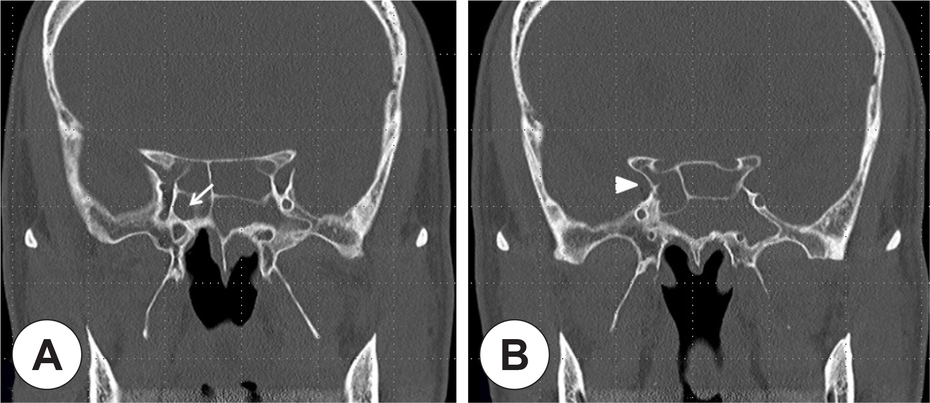
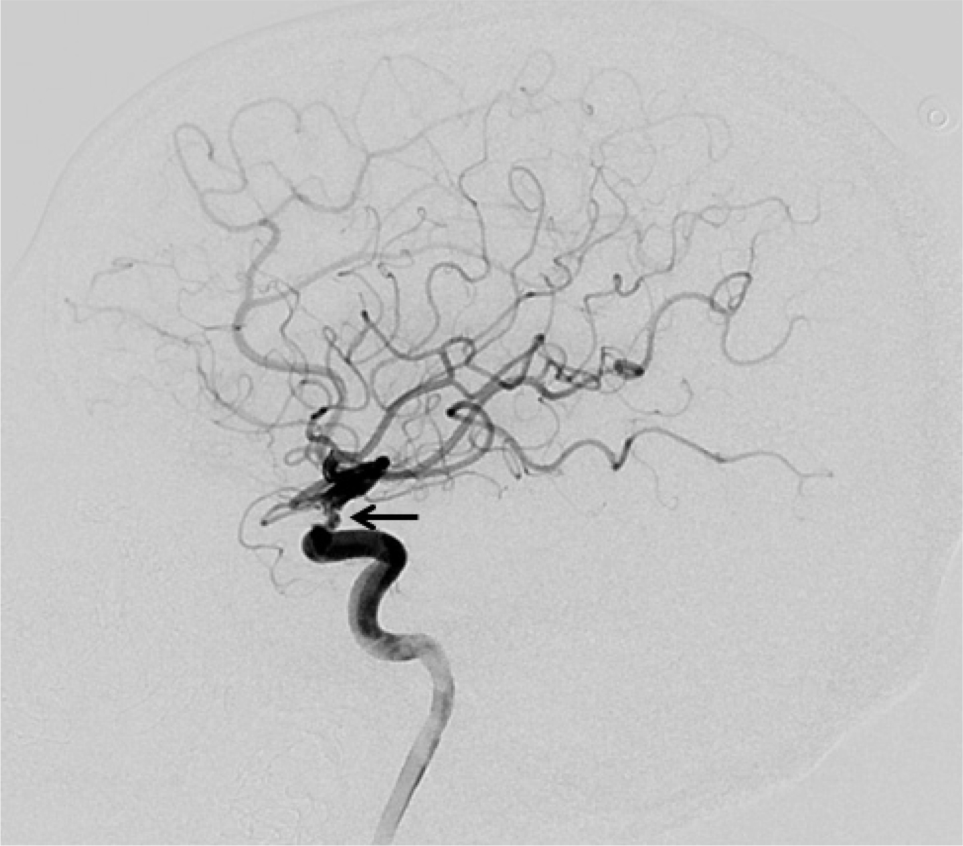
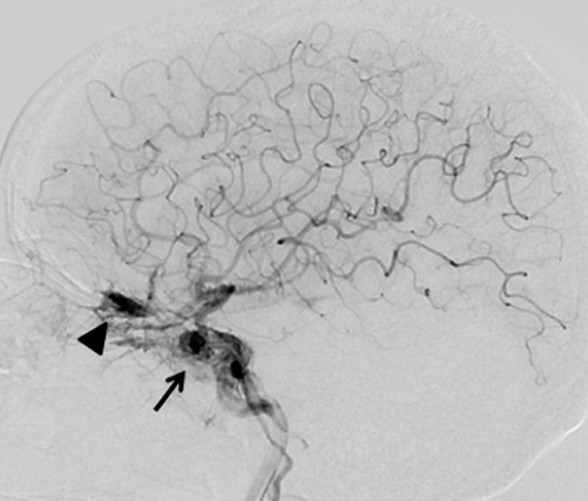
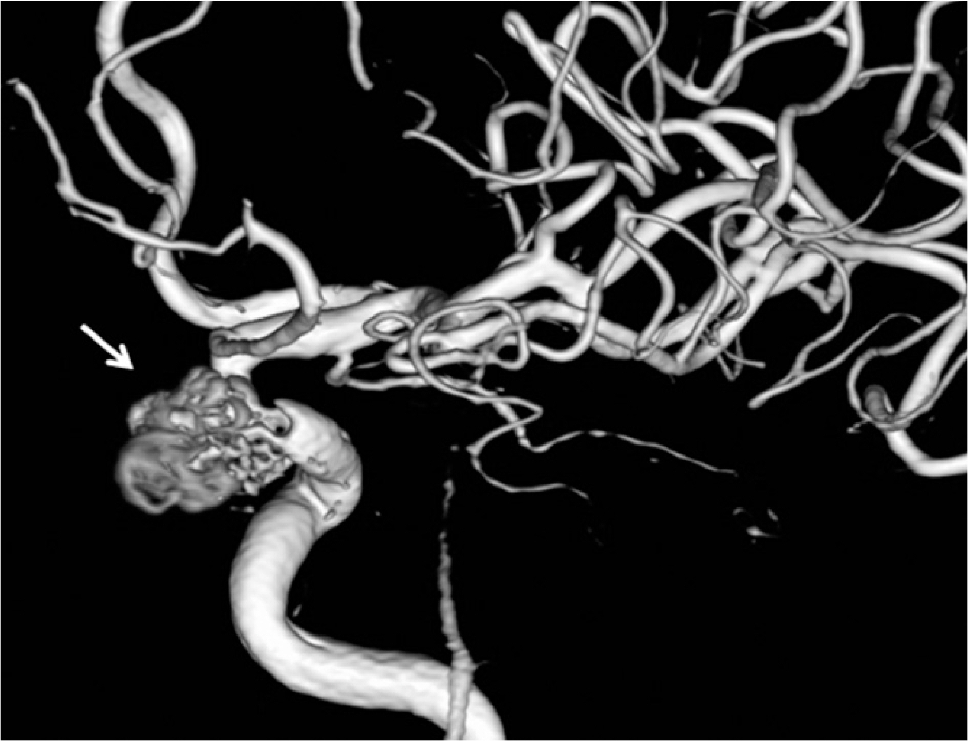
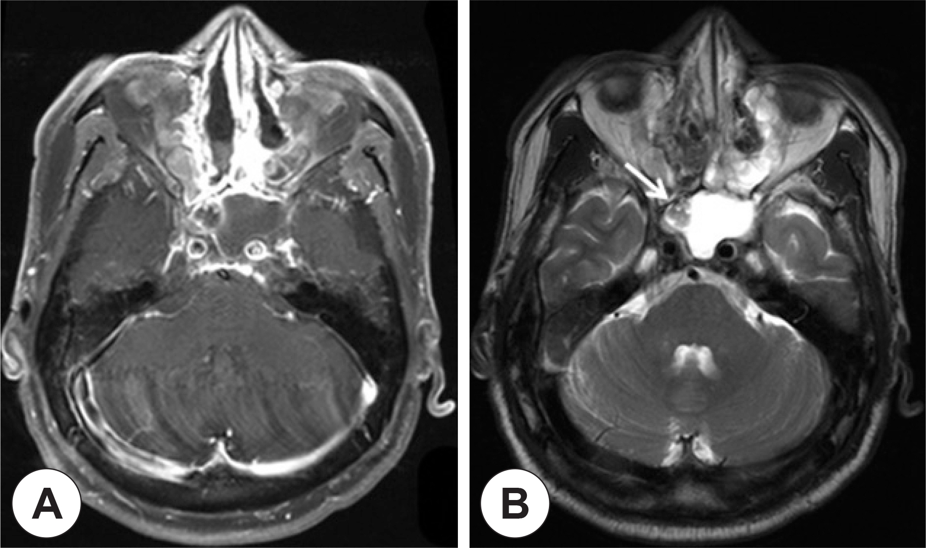
- Rupture of the internal carotid artery (ICA) during endoscopic sinus surgery is a rare complication. However, it can potentially result in death within minutes. In the event of a traumatic injury to the ICA during sphenoid sinus exploration, it is very difficult to control the bleeding. We present a case of carotid-cavernous fistula after an accidentally-developed ICA bleed during endoscopic sphenoidotomy. The patient was successfully treated with endovascular embolization techniques that included detachable microcoils.
MeSH Terms
Figure
Reference
-
References
1). May M, Levine HL, Mester SJ, Schaitkin B. Complication of endoscopic sinus surgery: Analysis of 2108 patients-incidence and prevention. Laryngoscope. 1994; 104(9):1080–3.2). Schaefer SD, Manning S, Close LG. Endoscopic paranasal sinus surgery: Indications and considerations. Laryngoscope. 1989; 99(1):1–5.3). Laws ER Jr. Vascular complications of transsphenoidal surgery. Pituitary. 1999; 2(2):163–70.4). Fujii K, Chambers SM, Rhoton AL Jr. Neurovascular relationships of the sphenoid sinus. A microsurgical study. J Neurosurg. 1979; 50(1):31–9.5). Lee SK, Park YS, Cho JH, Park YJ, Kang JM, Jeon EJ, et al. Anatomic variation of sphenoid sinus and related neurovascular structures: A study of CT analysis. Korean J Otolaryngol. 2004; 47:978–82.6). Sirikci A, Bayazit YA, Bayram M, Mumbuc S, Gungor K, Kan-likama M. Variations of sphenoid and related structures. Eur Radiol. 2000; 10(5):844–8.7). Kinnman J. Surgical aspects of the anatomy of the sphenoidal sinuses and the sella turcica. J Anat. 1977; 124:541–53.8). Elwany S, Yacout YM, Talaat M, El-Nahass M, Gunied A, Talaat M. Surgical anatomy of the sphenoid sinus. J Laryngol Otol. 1983; 97(3):227–41.
Article9). Kim HU, Kim SS, Kim IS, Chung IH, Yoon JH. Morphology of midline septum and accessory septum of the sphenoid sinus. Korean J Otolaryngol. 2001; 44(2):153–6.10). Raymod J, Hardy J, Czepko R, Roy D. Arterial injuries in transsphenoidal surgery for pituitary adenoma; the role of angiography and endovascular treatment. Am J Neuroradiol. 1997; 18(4):655–65.11). Lippert BM, Ringel K, Stoeter P, Hey O, Mann WJ. Stentgraft-implantation for treatment of internal carotid artery injury during endonasal sinus surgery. Am J Rhinol. 2007; 21(4):520–4.
Article12). 12) Valentine R, Wormald PJ. Carotid artery injury after endonasal surgery. Otolaryngol Clin North Am. 2011; 44(5):1059–79.13). Goodwin JR, Johnson MH. Carotid injury secondary to blunt head trauma: case report. J Trauma. 1994; 37(1):119–22.14). Barrow DL, Spector RH, Braun IF, Landman JA, Tindall SC, Tindall GT. Classification and treatment of spontaneous carotid-cav-ernous sinus fisulas. J Neurosurg. 1985; 62(2):248–56.15). Iida K, Uozumi T, Arita K, Nakahara T, Ohba S, Satoh H. Steal phenomenon in a traumatic carotid-carvernous fistula. J Trauma. 1995; 39(5):1015–7.16). Higashida RT, Halbach VV, Tsai FY, Norman D, Pribram HF, Mehringer CM, et al. Interventional neurovascular treatment of traumatic carotid and vertebral artery lesions: results in 234 cases. AJR Am J Roentgenol. 1989; 153(3):577–82.
Article17). Kim JK, Seo JJ, Kim YH, Kang HK, Lee JH. Traumatic bilateral cartotid-carvernous fistulas treated with detachable balloon. A case report. Acta Radiol. 1996; 37(1):46–8.18). Pierot L, Moret J, Boulin A, Castaings L. Endovascular treatment of posttraumatic complex carotid-cavernous fistulas, using the arterial approach. J Neuroradiol. 1992; 19(2):79–87.19). Halbach VV, Higashida RT, Barnwell SL, Dowd CF, Hieshima GB. Transarterial platinum coil embolization of cartotid-cavern-ous fistulas. Am J Neuroradiol. 1991; 12(3):429–33.20). Yang PJ, Halbach VV, Higashida RT, Hieshima GB. Platinum wire: a new transvascular embolic agent. Am J Neuroradiol. 1988; 9(3):547–50.
- Full Text Links
- Actions
-
Cited
- CITED
-
- Close
- Share
- Similar articles
-
- Direct Microsurgical Repair of Traumatic Carotid-Cavernous Fistula
- A Case of Dural Carotid-Cavernous Sinus Fistula Associated with Ophthalmic Manifestations
- Regional Cerebral Blood Flow Changes in Traumatic Carotid Cavernous Fistula During Trapping Procedure: Case Study, Preliminary Report
- Dural Carotid-Cavernous Sinus Fistula
- Parent artery occlusion of a giant internal carotid artery pseudoaneurysm-related direct carotid cavernous fistula: A case report

