J Rheum Dis.
2011 Mar;18(1):68-69. 10.4078/jrd.2011.18.1.68.
Chronic Cutaneous Lupus Erythematosus Presenting with a Buccal Erythema
- Affiliations
-
- 1Department of Dermatology, Hanyang University College of Medicine, Seoul, Korea. cwlee@hanyang.ac.kr
- KMID: 2223187
- DOI: http://doi.org/10.4078/jrd.2011.18.1.68
Abstract
- No abstract available.
MeSH Terms
Figure
-

Figure 1. A mucosal lesion of erythematous patch on the buccal mucosa is seen with ‘radiating white striae’ on the surface (open arrow).
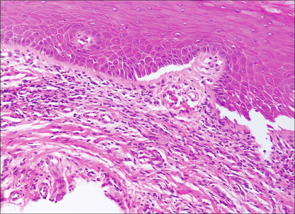
Figure 2. Biopsy from the mucosal lesion showed an interface dermatitis with foci of junctional clefts. A lymphocytic infiltration along the upper interstisium is apparent (H&E stain, ×200).
Reference
-
References
1. Rothfield N, Sontheimer RD, Bernstein M. Lupus erythematosus: systemic and cutaneous manifestations. Clin Dermatol. 2006; 24:348–62.2. Werth VP. Clinical manifestations of cutaneous lupus erythematosus. Autoimmun Rev. 2005; 4:296–302.3. Schi⊘dt M, Andersen L, Shear M, Smith CJ. Leukoplakia- like lesions developing in patients with oral discoid lupus erythematosus. Acta Odontol Scand. 1981; 39:209–16.4. Burge SM, Frith PA, Juniper RP, Wojnarowska F. Mucosal involvement in systemic and chronic cutaneous lupus erythematosus. Br J Dermatol. 1989; 121:727–41.
- Full Text Links
-
- Actions
-
Cited
- CITED
-
- Close
- Share
-
- Similar articles
-
- Two Cases of Hypertrophic Discoid Erythemas in Patients with Chronic Cutaneous Lupus Erythematosus
- Chronic Cutaneous Lupus Erythematosus With Hematologic/serologic Abnormalities: Incomplete Systemic Lupus Erythematosus
- A Case of Rowell's Syndrome
- A Case of Hypertrophic Lupus Erythematosus
- An Extensive Cutaneous Erythema associated with Vasculitis in a Patient with Systemic Lupus Erythematosus
