J Rheum Dis.
2011 Dec;18(4):327-328. 10.4078/jrd.2011.18.4.327.
Henoch-Schonlein Purpura with Hemorrhagic Bullous Skin Lesions
- Affiliations
-
- 1Division of Rheumatology, Department of Internal Medicine, Chonbuk National University Medical School and Research Institute of Clinical Medicine, Jeonju, Korea. ywhim@jbnu.ac.kr
- KMID: 2223141
- DOI: http://doi.org/10.4078/jrd.2011.18.4.327
Abstract
- No abstract available.
MeSH Terms
Figure
-
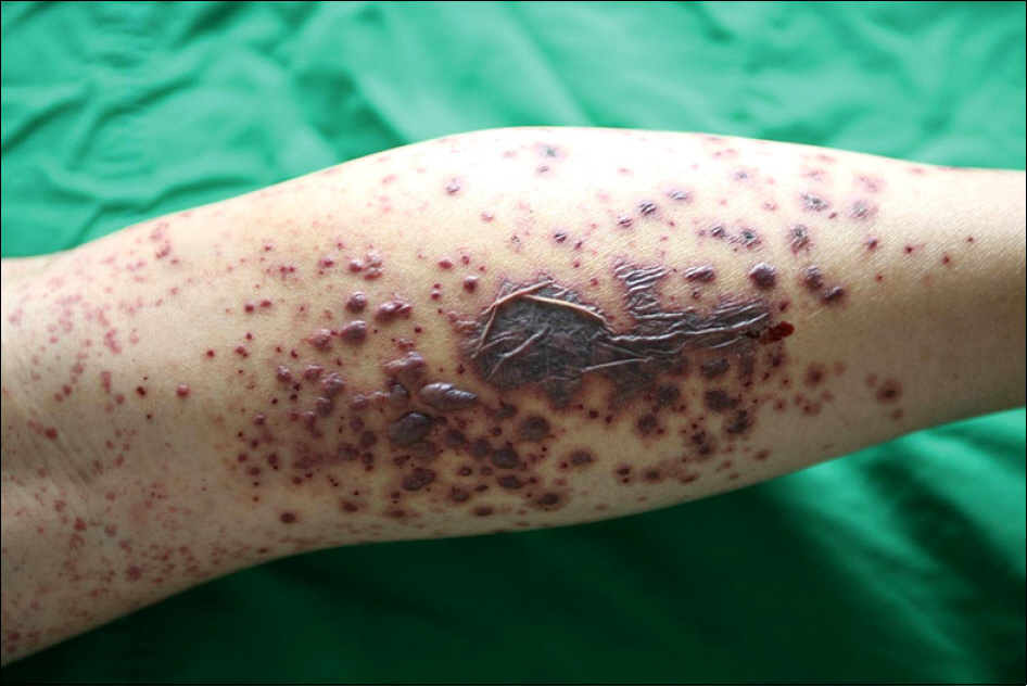
Figure 1. Multiple hemorrhagic bullous and purpuric lesions on the leg.
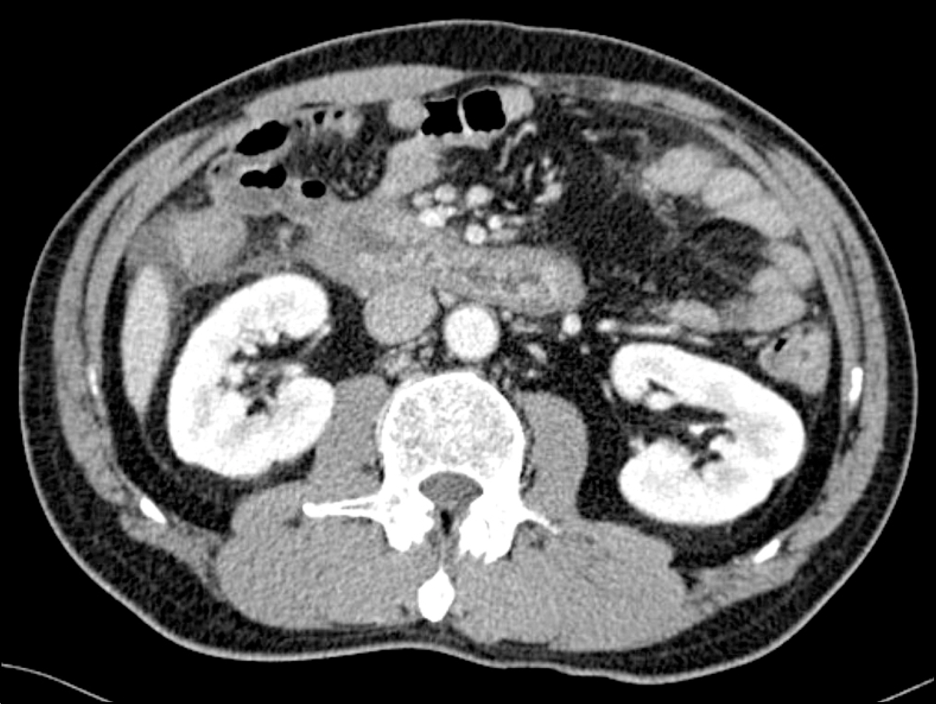
Figure 2. Edematous wall thickening of duodenal 3 rd portion and terminal ileum.
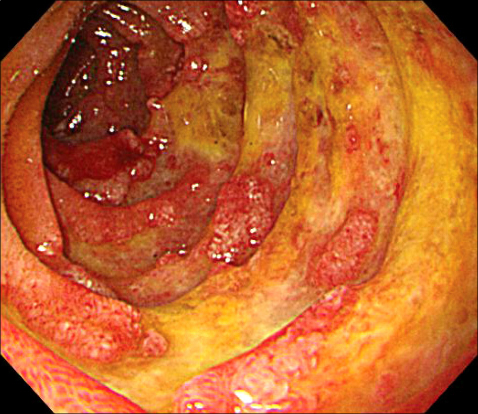
Figure 3. Several erythematous and edematous mucosa and pat-ches were seen at the bulb and the second portion of duodenum.

Figure 4. Biopsy from the bullae showed a necrotic epidermis, perivascular neutrophilic, lymphocytic infiltration with karyorr-hexis, and endothelial swelling, whichs was compatible with leukocytoclastic vasculitis.
Reference
-
References
1. Kausar S, Yalamanchili A. Management of haemorrhagic bullous lesions in Henoch-Schö nlein purpura: is there any consensus? J Dermatolog Treat. 2009; 20:88–90.2. Trapani S, Mariotti P, Resti M, Nappini L, de Martino M, Falcini F. Severe hemorrhagic bullous lesions in Henoch Schonlein purpura: three pediatric cases and review of the literature. Rheumatol Int. 2010; 30:1355–9.
Article3. Park SE, Lee JH. Haemorrhagic bullous lesions in a 3-year-old girl with Henoch-Schö lein purpura. Acta Paediatr. 2011; 100:e283–4.4. Shin JI, Lee JS. Hemorrhagic bullous lesions in Henoch-Schö nlein purpura. Pediatr Int. 2007; 49:121.
- Full Text Links
-
- Actions
-
Cited
- CITED
-
- Close
- Share
-
- Similar articles
-
- Two Cases of Henoch-Sch nlein Purpura Presenting as Bullous Lesions
- Hemorrhagic Bullous Lesions in a 9-year-old Girl with Henoch-Scholein Purpura
- A Case of Henoch-Schonlein Purpura Complicated by Hemorrhagic Ascites and Multiple Upper Gastrointestinal Bleeding
- A Case of Henoch-Schonlein Purpura with Hemorrhagic Bullous Lesions
- A Case of Hemorrhagic Bullous Henoch-Scholein Purpura: Cellulitis Presenting as a Complication
