Spontaneous Achilles Tendon Rupture in a Patient with Ankylosing Spondylitis
- Affiliations
-
- 1Department of Orthopedic Surgery, Daeun Hospital, Jeonju, Korea. oschae68@hanmail.net
- KMID: 2222792
- DOI: http://doi.org/10.4078/jrd.2016.23.2.136
Abstract
- No abstract available.
Figure
-
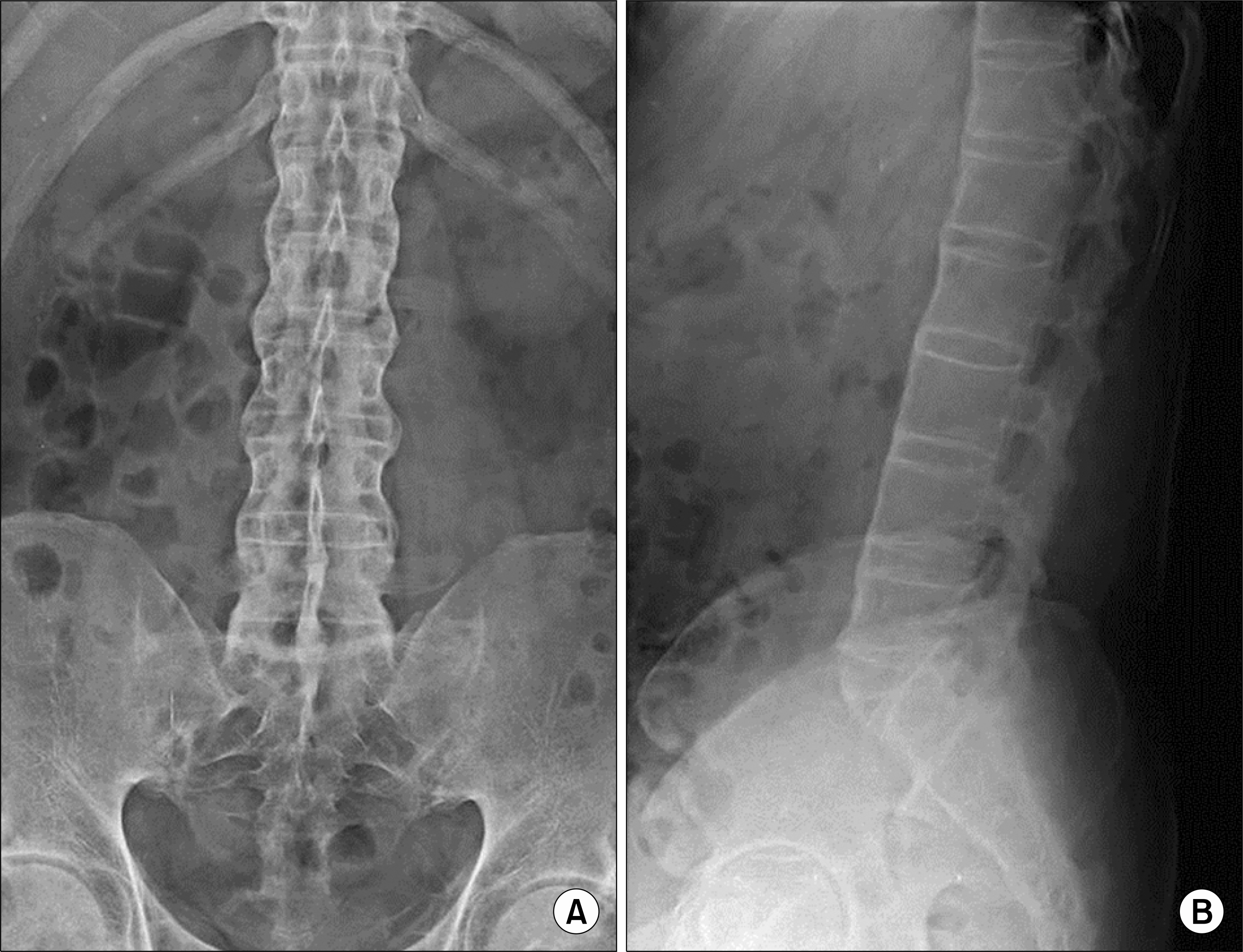
Figure 1. Simple anteroposterior (A) and lateral (B) L-spine radiography show a bamboo spine.
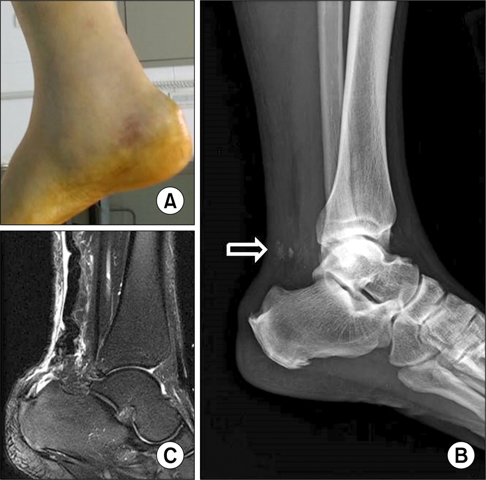
Figure 2. (A) The clinical photo shows swelling, ecchymosis and dimpling in the posterior aspect of the ankle. (B) Simple lateral ankle image shows loss of Kager's triangle and bony fragments (arrow). (C) Sagittal T2 magnetic resonance image shows rupture of the Achilles tendon at calcaneal insertion site and enthesopathic spur.
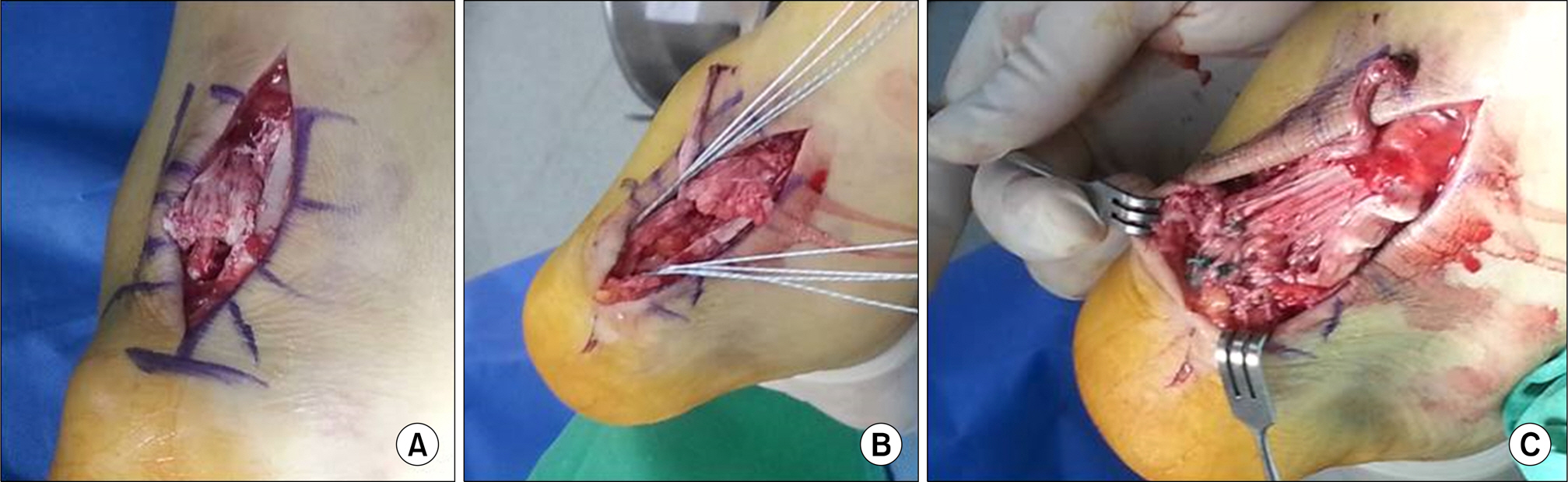
Figure 3. (A) Intraoperative finding shows the rupture of the Achilles tendon at calcaneal insertion site and combined with bony fragments. (B) Achilles tendon rupture was treated with tendon to bone repair using suture anchors. (C) Postoperative finding shows complete repair tendon to bone repair.
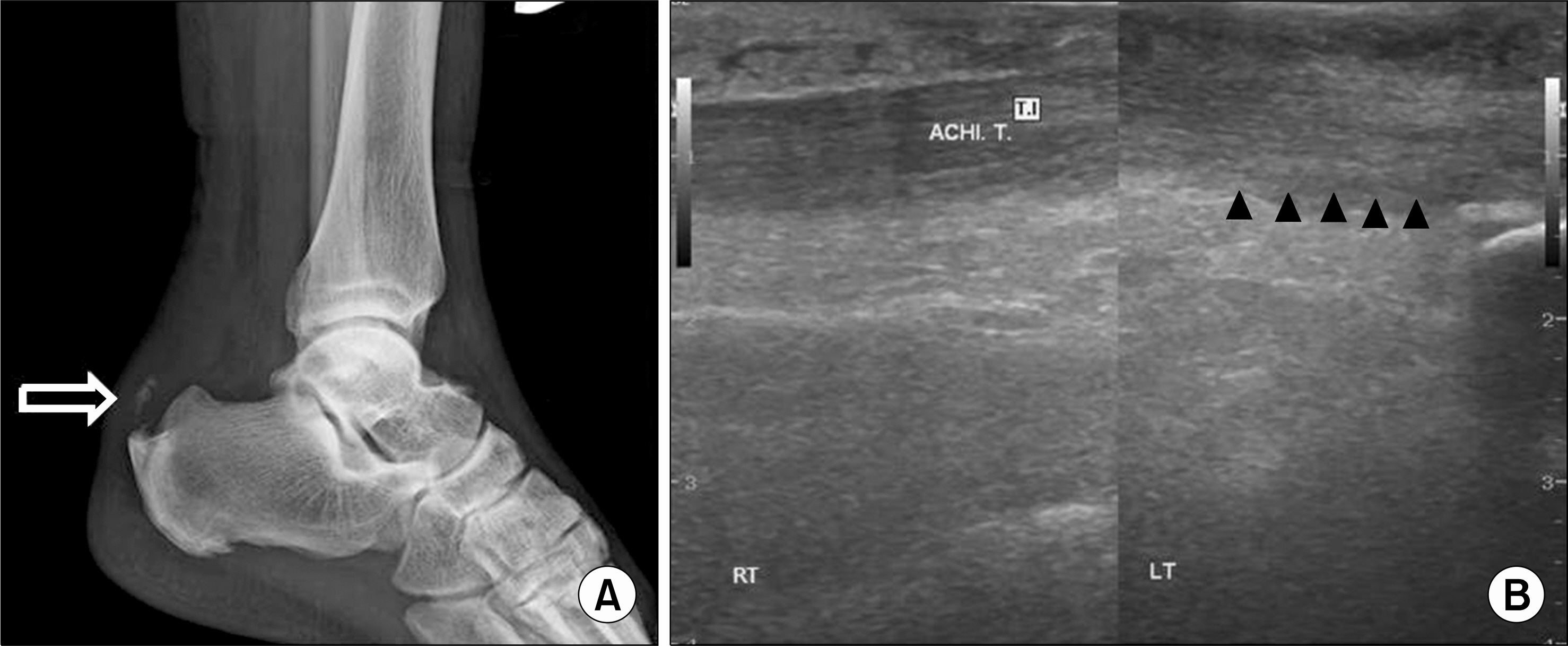
Figure 4. Six months follow-up simple lateral ankle image shows intact morphology of Kager's triangle (arrow) (A), ultrasonography shows normal distal Achilles tendon with fibrillar pattern at calcaneal insertion (arrowheads) (B). ACHI. T: Achilles tendon, LT: left, RT: right.
Reference
-
1. Braun J, Sieper J. Ankylosing spondylitis. Lancet. 2007; 369:1379–90.
Article2. Kim HW, Lee SH. Pathogenesis of ankylosing spondylitis. J Rheum Dis. 2015; 22:61–8.
Article3. Jiang N, Wang B, Chen A, Dong F, Yu B. Operative versus nonoperative treatment for acute Achilles tendon rupture: a meta-analysis based on current evidence. Int Orthop. 2012; 36:765–73.
Article
- Full Text Links
-
- Actions
-
Cited
- CITED
-
- Close
- Share
-
- Similar articles
-
- Spontaneous Achilles Tendon Rupture After Repeated Local Steroid Injention: A Case Report
- Spontaneous Bilateral Achilles Tendon Rupture after Local Steroid Injection for Carpal Tunnel Syndrome in a Diabetic Patient: A case report
- Achilles Tendon Rupture Associated With Ipsilateral Medial Malleolar Fracture (A Case Report)
- Reconstruction of Chronic Achilles Tendon Rupture Using Interposed Scar Tissue (A Report of Two Cases)
- Heterotopic Ossification of a Partially Ruptured Achilles Tendon (A Case Report)
