J Rheum Dis.
2013 Jun;20(3):202-203. 10.4078/jrd.2013.20.3.202.
Dysphagia Caused by Ossificaion of the Cervical Anterior Longitudinal Ligament
- Affiliations
-
- 1Department of Neurosurgery, Chosun University Hospital, Gwangju, Korea.
- 2Department of Internal Medicine, Soonchunhyang University Seoul Hospital, Seoul, Korea. healthyra@schmc.ac.kr
- KMID: 2222763
- DOI: http://doi.org/10.4078/jrd.2013.20.3.202
Abstract
- No abstract available.
MeSH Terms
Figure
-
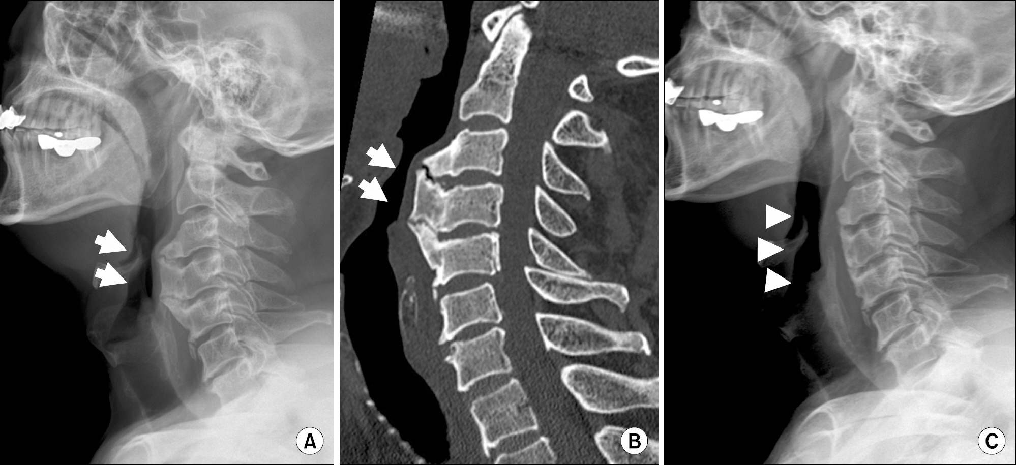
Figure 1. (A) It shows ossification of the anterior longitudinal ligament (OALL) from C3 to C5 (arrows). (B) Cervical computed tomography image shows compression of trachea by OALL (arrows). (C) Postoperative cervical radiograph shows removal of OALL which have compressed the esophagus and trachea (arrow heads).
Reference
-
References
1. Lecerf P, Malard O. How to diagnose and treat sympto-matic anterior cervical osteophytes? Eur Ann Otorhinolar-yngol Head Neck Dis. 2010; 127:111–6.
Article2. Epstein NE, Hollingsworth R. Ossification of the cervical anteriorlongitudinal ligament contributing to dysphagia. Case report. J Neurosurg. 1999; 90(2 Suppl):261–3.
- Full Text Links
-
- Actions
-
Cited
- CITED
-
- Close
- Share
-
- Similar articles
-
- Dysphagia Caused by Ossification of the Cervical Anterior Longitudinal Ligament
- Dysphagia Caused by Ossificaion of the Cervical Anterior Longitudial Ligament : Report of Two Cases
- Ossification of the Posterior Longitudinal Ligament: 2 cases report
- Treatment of Ossification of Posterior Longitudinal Ligament in Cervical Spine with Anterior Fusion and Anterior Decompression: Report of 3 Cases
- Improvement of Dysphagia after Anterior Cervical Screw Removal: Case Report
