Clin Endosc.
2015 Jan;48(1):81-84. 10.5946/ce.2015.48.1.81.
Colonic Lymphangiomatosis Resolved after Excisional Biopsy
- Affiliations
-
- 1Department of Internal Medicine, Sahmyook Medical Center, Seoul, Korea.
- 2Department of Pathology, Sahmyook Medical Center, Seoul, Korea.
- 3Division of Gastroenterology, Department of Internal Medicine, Konkuk University Medical Center, Konkuk University School of Medicine, Seoul, Korea. chansshim@naver.com
- KMID: 2221765
- DOI: http://doi.org/10.5946/ce.2015.48.1.81
Abstract
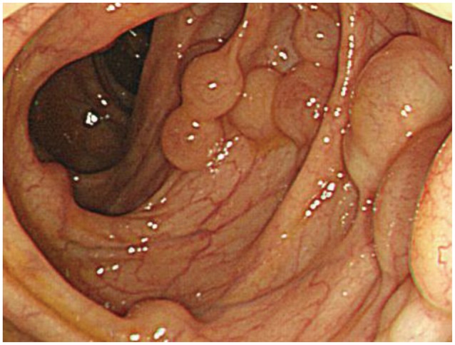
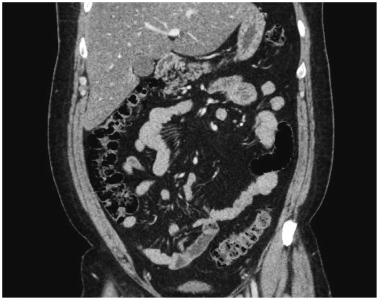
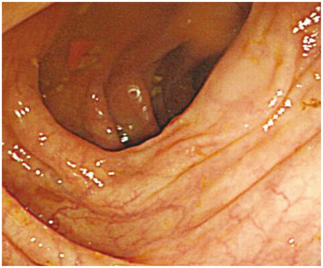
- Lymphangioma is an uncommon malformation of the lymphatic system that involves a benign proliferation of the lymphatics, with no established treatment method. Multiple colonic lymphangioma, or colonic lymphangiomatosis, is an extremely rare condition. We report a case of colonic lymphangiomatosis that was detected during a colonoscopic examination conducted as part of a general health check-up. The lesion completely resolved after excisional biopsy.
Keyword
MeSH Terms
Figure
Reference
-
1. Watanabe T, Kato K, Sugitani M, et al. A case of multiple lymphangiomas of the colon suggesting colonic lymphangiomatosis. Gastrointest Endosc. 2000; 52:781–784. PMID: 11115919.
Article2. Pipinos II, Baxter BT. The lymphatics. In : Townsend CM, Beauchamp RD, Evers BM, editors. Textbook of Surgery: the Biological Basis of Modern Surgical Practice. 17th ed. Philadelphia: W.B. Saunders;2004. p. 2078.3. Jung SW, Cha JM, Lee JI, et al. A case report with lymphangiomatosis of the colon. J Korean Med Sci. 2010; 25:155–158. PMID: 20052363.
Article4. Lee JM, Chung WC, Lee KM, et al. Spontaneous resolution of multiple lymphangiomas of the colon: a case report. World J Gastroenterol. 2011; 17:1515–1518. PMID: 21472113.
Article5. Kochman ML, Wiersema MJ, Hawes RH, Canal D, Wiersema L. Preoperative diagnosis of cystic lymphangioma of the colon by endoscopic ultrasound. Gastrointest Endosc. 1997; 45:204–206. PMID: 9041015.
Article6. Matsuda T, Matsutani T, Tsuchiya Y, et al. A clinical evaluation of lymphangioma of the large intestine: a case presentation of lymphangioma of the descending colon and a review of 279 Japanese cases. J Nippon Med Sch. 2001; 68:262–265. PMID: 11404774.
Article7. Pusztaszeri MP, Seelentag W, Bosman FT. Immunohistochemical expression of endothelial markers CD31, CD34, von Willebrand factor, and Fli-1 in normal human tissues. J Histochem Cytochem. 2006; 54:385–395. PMID: 16234507.
Article8. Fukunaga M. Expression of D2-40 in lymphatic endothelium of normal tissues and in vascular tumours. Histopathology. 2005; 46:396–402. PMID: 15810951.
Article9. Enzinger FM, Weiss SW. Tumors of lymph vessels. In : Enzinger FM, Weiss SW, editors. Soft Tissue Tumors. 3rd ed. St. Louis: Mosby;1995. p. 679–699.
- Full Text Links
- Actions
-
Cited
- CITED
-
- Close
- Share
- Similar articles
-
- Colonic Lymphangiomatosis with Normal Colonoscopic Finding in an Adult
- A Case Report with Lymphangiomatosis of the Colon
- Metachronous Bilateral Renal Lymphangiomatosis Mimicking as a Simple Renal Cyst
- Lymphangiomatosis of Bone and Soft Tissue: A Case Report
- Congenital lymphangiomatosis of the right lower limb


